Michael Pridmore1, Maria Garza1, Laura Jones2, Cassandra Reynolds2, Deepak Gupta2, Manus Donahue1, and Rachelle Crescenzi1
1Radiology and Radiological Sciences, Vanderbilt University Medical Center, Nashville, TN, United States, 2Cardiovascular Medicine, Vanderbilt University Medical Center, Nashville, TN, United States
1Radiology and Radiological Sciences, Vanderbilt University Medical Center, Nashville, TN, United States, 2Cardiovascular Medicine, Vanderbilt University Medical Center, Nashville, TN, United States
Free-breathing pCASL MRI measuring cortical kidney RBF shows promise for applications in clinical research. SSBP showed reduced renal blood flow, consistent with poor sodium handling, which may provide a noninvasive clinically feasible imaging biomarker for salt sensitivity.
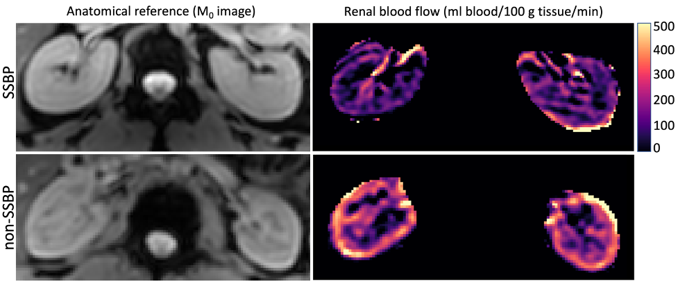
Figure 4. Case examples of renal blood flow maps in persons A) with SSBP and B) non-SSBP. Cortical renal blood flow maps (ml blood/100 g tissue/min) are shown from the 4x acquisition protocol. The subject with SSBP exhibits lower cortical perfusion compared to the non-SSBP subject, consistent with group trends. The color bar and range of values for renal blood flow are shown top-right using a perceptually uniform colormap (magma)7.
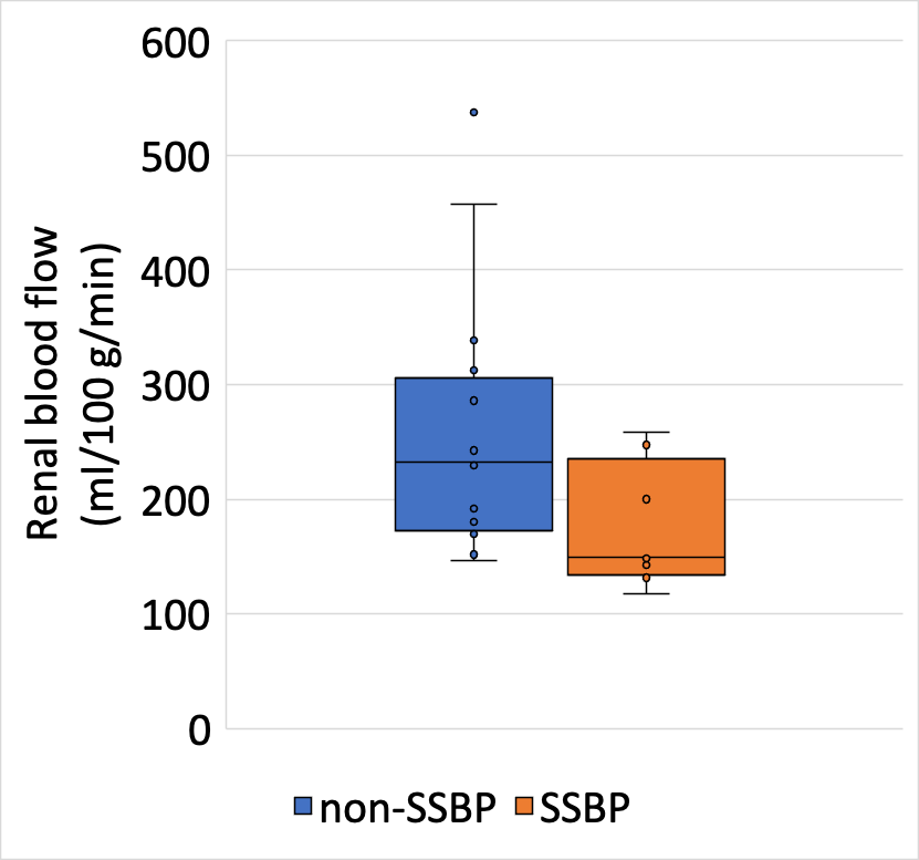
Figure 3. Renal blood flow (RBF) in groups with SSBP and non-SSBP. Salt sensitive blood pressure (SSBP) and non-SSBP groups were compared for cortical RBF, that demonstrated a trend towards reduced cortical RBF in SSBP (174.5 ± 53.9 mL/100g/min) compared to non-SSBP (253.8 ± 111.7 mL/100g/min) groups (p=0.07). This box plot demonstrates the median as the central line, and upper and lower quartiles as the edges of the boxes.
