Yiming Wang1, Limin Zhou1, Ivan Pedrosa1,2,3, and Ananth J. Madhuranthakam1,2
1Radiology, UT Southwestern Medical Center, Dallas, TX, United States, 2Advanced Imaging Research Center, UT Southwestern Medical Center, Dallas, TX, United States, 3Urology, UT Southwestern Medical Center, Dallas, TX, United States
1Radiology, UT Southwestern Medical Center, Dallas, TX, United States, 2Advanced Imaging Research Center, UT Southwestern Medical Center, Dallas, TX, United States, 3Urology, UT Southwestern Medical Center, Dallas, TX, United States
We applied a variable density sampling method to renal
ASL, which acquires the center of the k-space with higher averages and improves
SNR and robustness, and combined it with partial k-space acquired M0 to
compensate for increased scan time, but without compromising perfusion
quantification
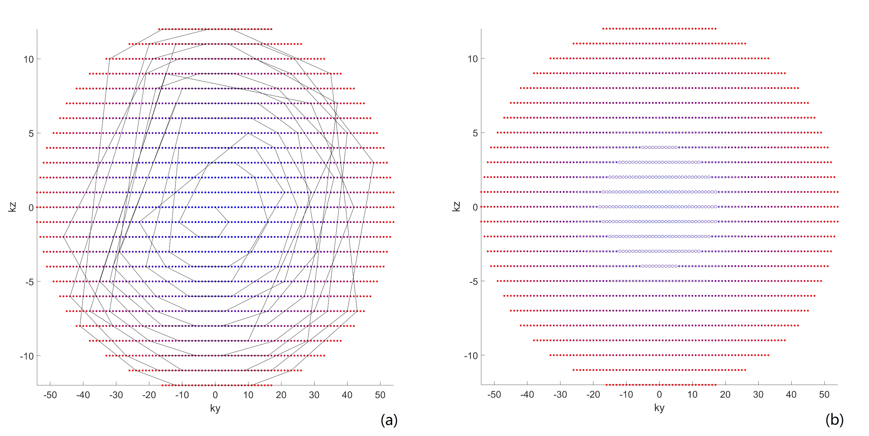
Figure
1. (a) A Cartesian grid of a ky-kz
space showing CASPR view ordering, where the earlier echoes are acquired at the
beginning of each echo train (blue dots) following a pseudo-spiral trajectory
towards the later echoes (red). (b) VD-CASPR method acquires profiles in region
1 (R1, open circle), 2 (R2, asteroid) and R3 (dot) with variable density (e.g.
3, 2, and 1 averages respectively), but still maintaining a spiral profile
ordering on a Cartesian grid for each echo train.
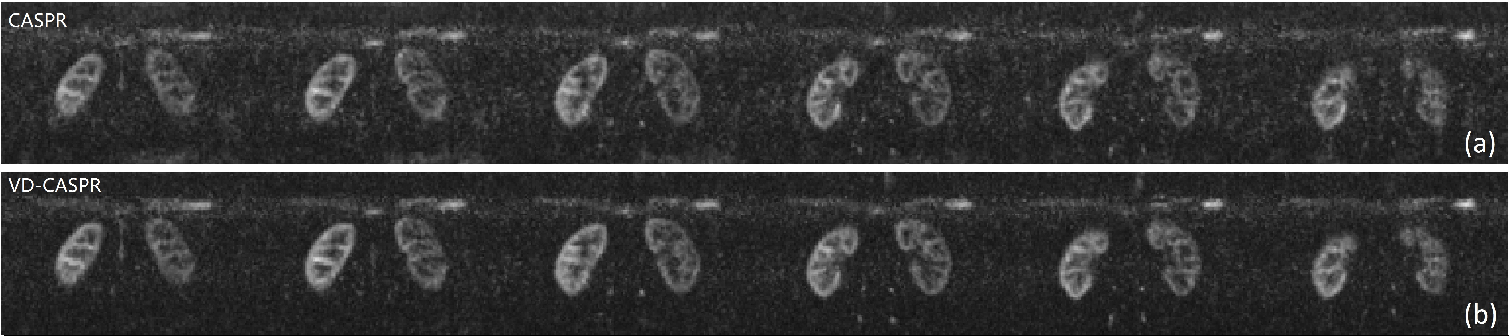
Figure 2. Kidney perfusion
weighted images of a normal volunteer acquired using single-average 3D TSE-CASPR
(a, top row) and with VD-CASPR method (b, bottom row), shown for several slices
of the kidneys. VD-CASPR images showed minimized background noise and improved
SNR. Note some signal variation between the right and left kidneys, probably
due to B1 inhomogeneities.