A. Max Hamilton1,2,3,4, Qandeel Shafqat1,2,3,4, Nils D. Forkert1,2,3, Ying Wu1,2,3,4, and Jeff F. Dunn1,2,3,4
1Department of Radiology, University of Calgary, Calgary, AB, Canada, 2Hotchkiss Brain Institute, University of Calgary, Calgary, AB, Canada, 3Department of Clinical Neurosciences, University of Calgary, Calgary, AB, Canada, 4Experimental Imaging Center Cumming School of Medicine, University of Calgary, Calgary, AB, Canada
1Department of Radiology, University of Calgary, Calgary, AB, Canada, 2Hotchkiss Brain Institute, University of Calgary, Calgary, AB, Canada, 3Department of Clinical Neurosciences, University of Calgary, Calgary, AB, Canada, 4Experimental Imaging Center Cumming School of Medicine, University of Calgary, Calgary, AB, Canada
Using 9.4T MRI, a cryoprobe, and atlas-based volumetric analysis, we
identified subcortical and corpus callosum atrophy in the cuprizone mouse model
of MS, following chronic demyelination at 12 weeks.
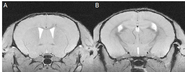
Figure 1. Corpus
callosum demyelination in cuprizone mice. Demyelination follows a rostro-caudal
pattern. A) Lateral callosal demyelination rostrally. B) Medial callosal
demyelination caudally.
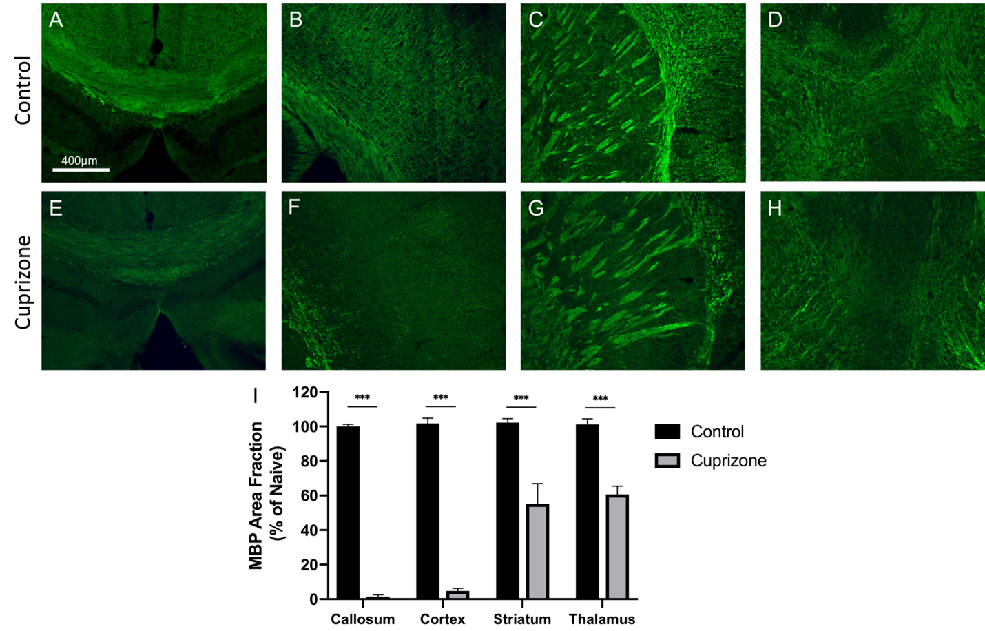
Figure 4. Cuprizone mice experience severe
demyelination after 12 weeks on cuprizone diet compared to controls (reduced
green fluorescence). Near complete demyelination seen in medial corpus callosum
(A, E) and cortex (B, F). Partial demyelination was seen in the striatum (C, G)
and the thalamus (D, H). Statistics were performed using Student's t-test (I). p < 0.05*;
<0.01**, <0.001***.