Gustavo Chau Loo Kung1, Juliet Knowles2, Lijun Ni2, John Huguenard2, Michelle Monje2, and Jennifer McNab3
1Bioengineering Department, Stanford University, Stanford, CA, United States, 2Neurology Department, Stanford University, Stanford, CA, United States, 3Radiology Department, Stanford University, Stanford, CA, United States
1Bioengineering Department, Stanford University, Stanford, CA, United States, 2Neurology Department, Stanford University, Stanford, CA, United States, 3Radiology Department, Stanford University, Stanford, CA, United States
Our results on ex vivo mouse brains suggest a decrease in MRI derived g-ratios in the genu of the corpus callosum of a mouse model of absence epilepsy, in accordance with a similar decrease on ground truth EM measurements.
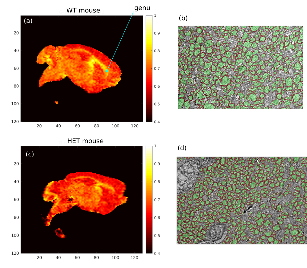
Figure 1. Sample images showing the MRI-derived g-ratio maps for a WT mouse (a) and a HET mouse (c). The genu region is indicated in (a). EM images with 1000x zoom and segmentation of myelin (red) and axons (green) for the same two mice are shown in (b) and (d) for the WT and HET mice respectively.
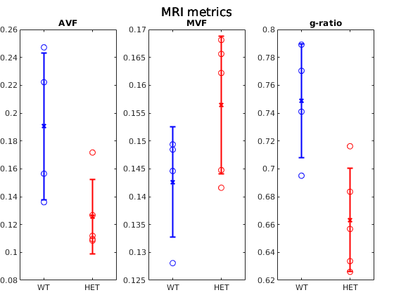
Figure 3. Error and scatter plot showing the MRI measurements of AVF, MVF and g-ratio of the WT mice (blue) and HET mice (red).