Joseph H. C. Lai1, Jiaxin Liu2, Jianpan Huang1, Yang Liu1, Zilin Chen1, Peng Xiao1, Gilberto K. K. Leung2, and Kannie W. Y. Chan1,3,4
1Department of Biomedical Engineering, City University of Hong Kong, Hong Kong, Hong Kong, 2Division of Neurosurgery, Department of Surgery, Li Ka Shing Faculty of Medicine, The University of Hong Kong, Hong Kong, Hong Kong, 3Russell H. Morgan Department of Radiology and Radiological Science, Johns Hopkins University School of Medicine, Baltimore, MD, United States, 4City University of Hong Kong Shenzhen Research Institute, Shenzhen, China
1Department of Biomedical Engineering, City University of Hong Kong, Hong Kong, Hong Kong, 2Division of Neurosurgery, Department of Surgery, Li Ka Shing Faculty of Medicine, The University of Hong Kong, Hong Kong, Hong Kong, 3Russell H. Morgan Department of Radiology and Radiological Science, Johns Hopkins University School of Medicine, Baltimore, MD, United States, 4City University of Hong Kong Shenzhen Research Institute, Shenzhen, China
This study examined the feasibility of CEST in monitoring ICH and its progression over two weeks. Decrease in rNOE and APT signals in lesion were observed on day7 and day3, respectively, when compared to contralateral side under insignificant iron suppression effect.
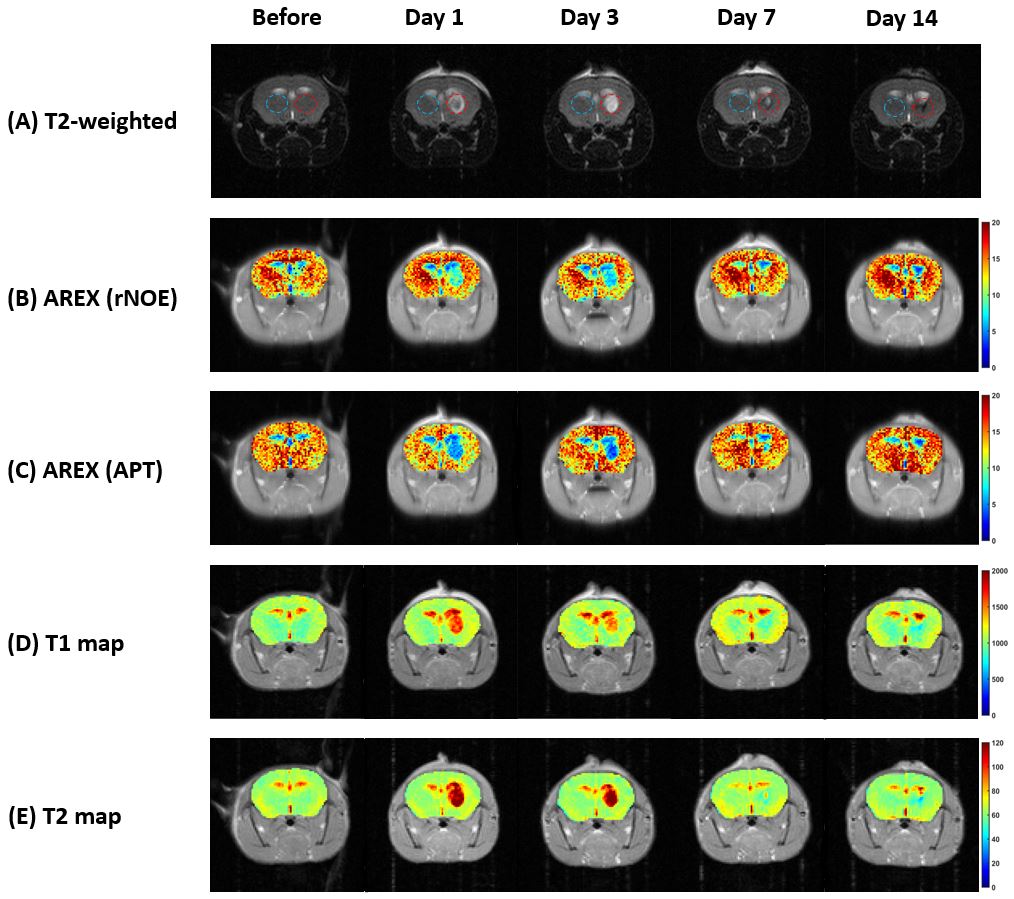
MR images of an ICH mouse at different time points (From
left to right: before, day 1, day 3, day 7, day 14) Pixels values of the
ipsilateral and the contralateral brain were extracted from the regions within
the red and blue circles respectively. (A)
T2-weighted reference image, (B)
AREX(rNOE), (C) AREX(APT), (D) T1 map, (E) T2 map.
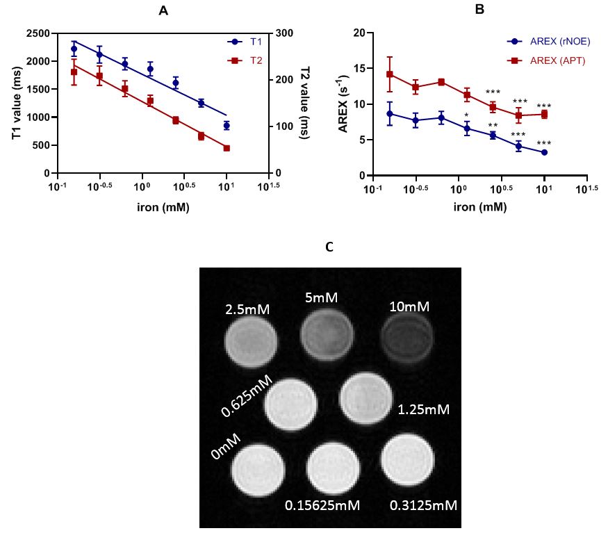
(A) T1 and T2
fitting with iron content (R2 for T1 = 0.9316, Y = -734.4*log X +
1769; R2 for T2 = 0.9792, Y = -96.96*log X + 153.6). (B) AREX value with iron content.
Two-way ANOVA was performed for statistical analysis as compare with the value
of 0.15625mM. *P<0.05, **P<0.01, ***P<0.001. (C) A reference T2-weighted image of the phantom. Values were taken
by averaging the pixels values of each tube.