Yaewon Kim1, Hsin-Yu Chen1, Adam W. Autry1, Javier Villanueva-Meyer1, Susan M. Chang2, Yan Li1, Peder E. Z. Larson1, Jeffrey R. Brender3, Murali C. Krishna3, Duan Xu1, Daniel B. Vigneron1,2, and Jeremy W. Gordon1
1Department of Radiology and Biomedical Imaging, University of California, San Francisco, CA, United States, 2Department of Neurological Surgery, University of California, San Francisco, CA, United States, 3Center for Cancer Research, National Cancer Institute, National Institutes of Health, Bethesda, MD, United States
1Department of Radiology and Biomedical Imaging, University of California, San Francisco, CA, United States, 2Department of Neurological Surgery, University of California, San Francisco, CA, United States, 3Center for Cancer Research, National Cancer Institute, National Institutes of Health, Bethesda, MD, United States
A new patch-based denoising approach using singular value decomposition for dynamic hyperpolarized [1-13C]pyruvate EPI scans of the human brain demonstrated dramatic improvements in image quality and quantification of metabolite dynamics.
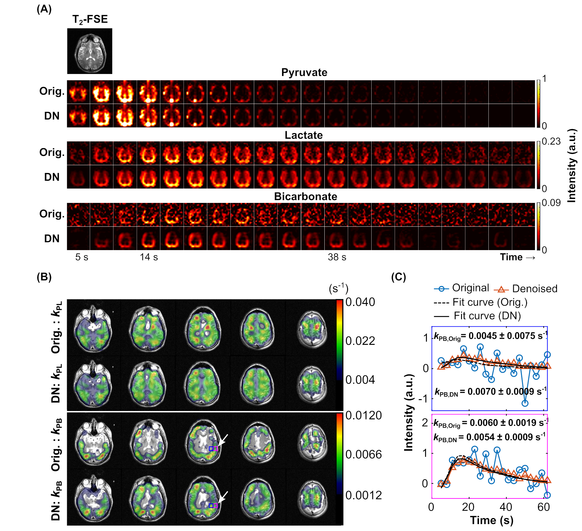
Figure 4. (A) Dynamic HP-13C EPI data of [1-13C]pyruvate, [1-13C]lactate, and [13C]bicarbonate signals from a healthy brain volunteer before (‘Orig.') and after applying GL-HOSVD (‘DN’). (B) Pyruvate-to-lactate (kPL) and pyruvate-to-bicarbonate (kPB) conversion rate maps before and after applying GL-HOSVD. (C) Dynamic traces of the original and denoised bicarbonate signals from 2 selected voxels, indicated by an arrow in the kPB maps, and the fitted kPB values. The denoised images show a 2-fold increase in spatial coverage of kPB maps with acceptable fit quality.
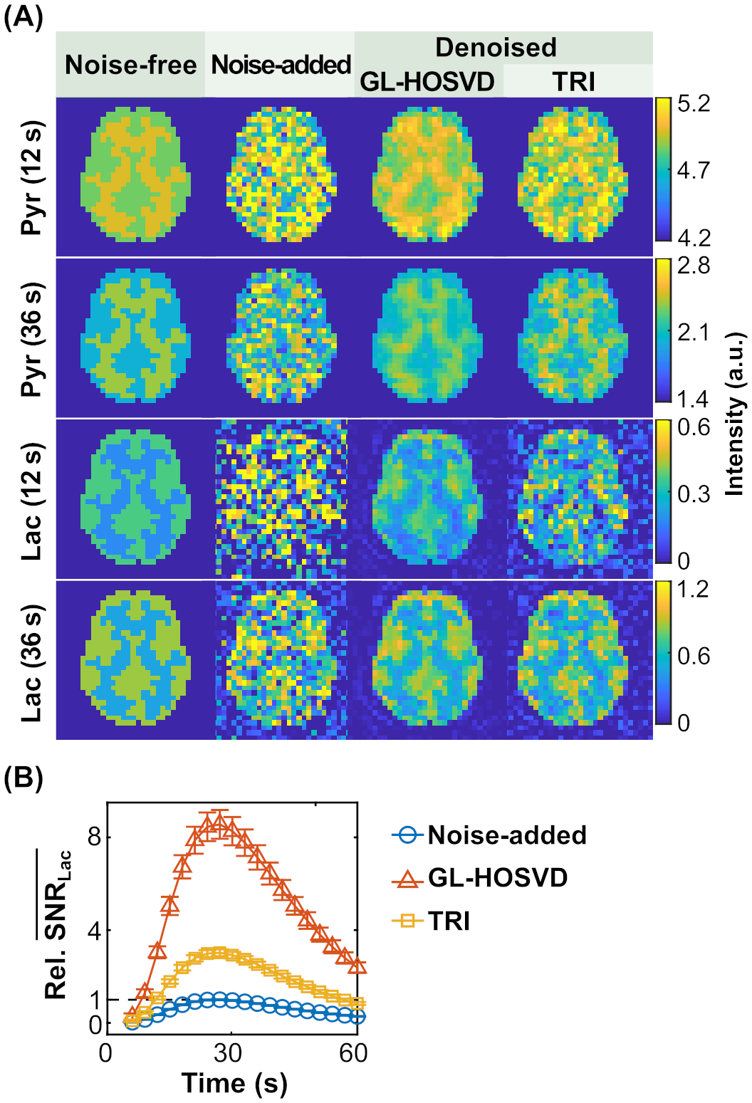
Figure 2. Comparative results between GL-HOSVD and Tensor Rank Truncation-Image Enhancement (TRI) methods for denoising simulated image data. (A) Pyruvate and lactate images at an early (t = 12 s) and late (t = 36 s) timepoints before and after denoising. (B) Relative mean values of signal-to-noise ratios of the noise-added and denoised lactate signals from 500 simulated datasets, showing 2 - 3 times higher SNR gain by using GL-HOSVD versus TRI.