Andrew Palmera Leynes1,2, Nikou Louise Damestani3, David John Lythgoe3, Ana Beatriz Solana4, Brice Fernandez5, Brian Burns1,6, Steven Charles Rees Williams3, Fernando Zelaya3, Peder E.Z. Larson1,2, and Florian Wiesinger3,4
1Department of Radiology and Biomedical Imaging, University of California San Francisco, San Francisco, CA, United States, 2UC Berkeley - UC San Francisco Joint Graduate Program in Bioengineering, Berkeley and San Francisco, CA, United States, 3King's College London, London, United Kingdom, 4GE Healthcare, Munich, Germany, 5GE Healthcare, Paris, France, 6GE Healthcare, Menlo Park, CA, United States
1Department of Radiology and Biomedical Imaging, University of California San Francisco, San Francisco, CA, United States, 2UC Berkeley - UC San Francisco Joint Graduate Program in Bioengineering, Berkeley and San Francisco, CA, United States, 3King's College London, London, United Kingdom, 4GE Healthcare, Munich, Germany, 5GE Healthcare, Paris, France, 6GE Healthcare, Menlo Park, CA, United States
We introduced significant improvements to the spatial and temporal
resolution of Looping Star using the “extreme MRI” approach, demonstrated
across two fMRI tasks.
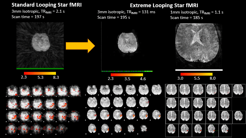
Animated Figure 3. Standard
Looping Star fMRI (left) vs high-temporal resolution (middle) and high-spatial
resolution (right) extreme Looping Star fMRI. Performing fMRI with a motor task can be done with either higher temporal resolution
(middle) or higher spatial resolution (right) that preserves structural detail
compared to standard Looping Star (left). An animated GIF (5x speedup) of the
temporal volumes is shown (top row) and the corresponding thresholded motor
task activation maps (bottom row) analyzed using FSL FEAT.
Only 1 second is shown due to file size limits.
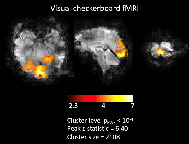
Figure 5. Demonstration of Extreme
Looping Star for a visual task at a different site. The activation map clearly
shows activation in the visual cortex at a TR of 0.155s and 1280 volumes with
an effective sub-Nyquist sampling factor per volume of 0.025. This is 10x finer temporal resolution
than original Looping Star.