Wiktor Olszowy1,2 and Ileana O Jelescu1,2
1CIBM Center for Biomedical Imaging, Lausanne, Switzerland, 2Animal Imaging and Technology, EPFL, Lausanne, Switzerland
1CIBM Center for Biomedical Imaging, Lausanne, Switzerland, 2Animal Imaging and Technology, EPFL, Lausanne, Switzerland
We observed task-induced water diffusivity decreases in the perfusion-free b-value regime. Moreover, we found that positive correlations were largely preserved while anti-correlations were suppressed in dfMRI compared to BOLD. We conclude that dfMRI is distinct from BOLD mechanisms.
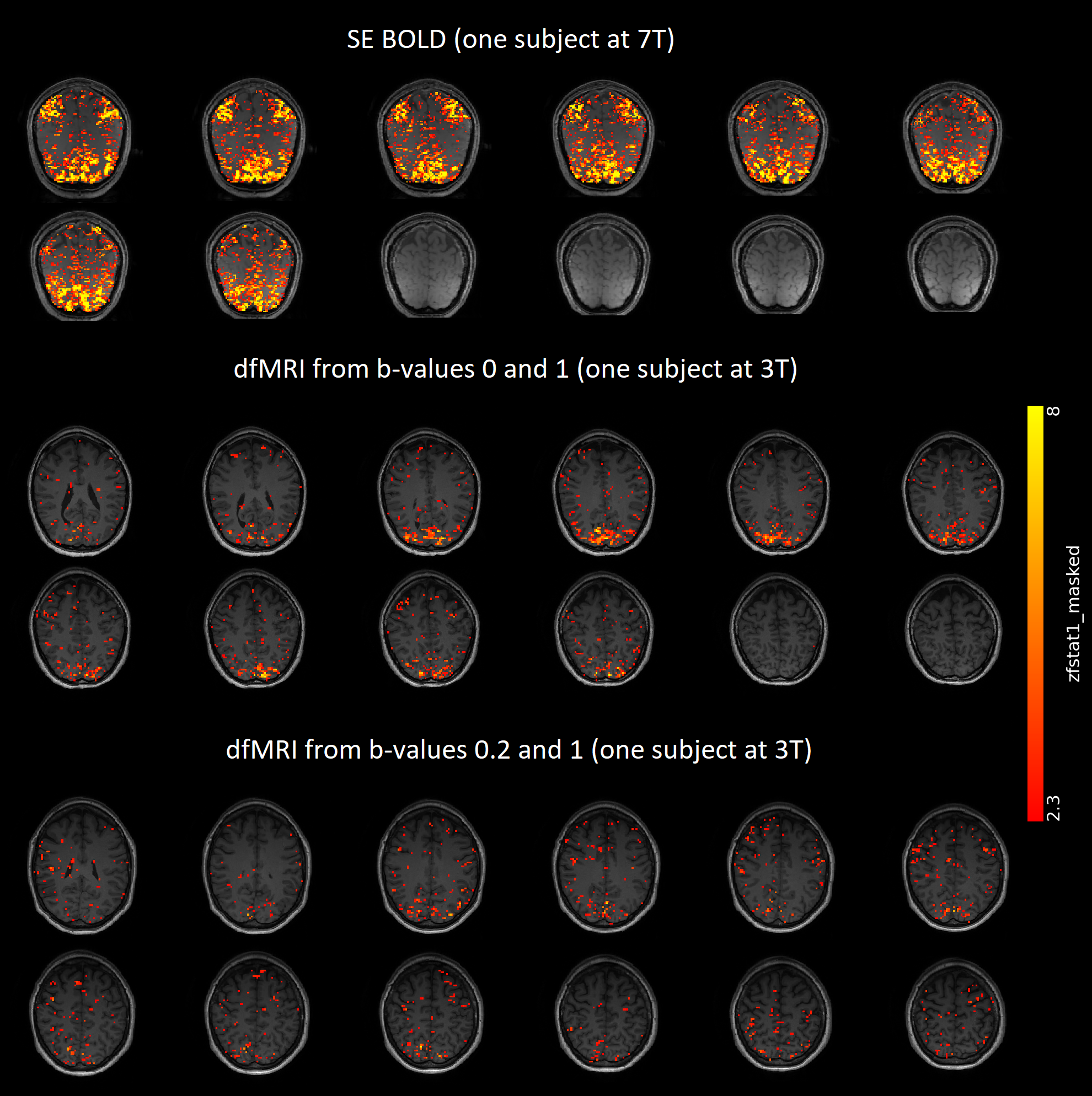
Fig 2: Whole-brain single-subject GLM results for SE
BOLD and dfMRI. In order to find activation without making assumptions about
the shape of the response functions, a Finite Impulse Response (FIR) set was
used. Significance was assessed with F-tests on the FIR regressors. The
F-statistic maps were converted to the displayed z-statistic maps, which were
additionally truncated at 2.3. Activation in visual and motor cortices was
observed for all three modalities.
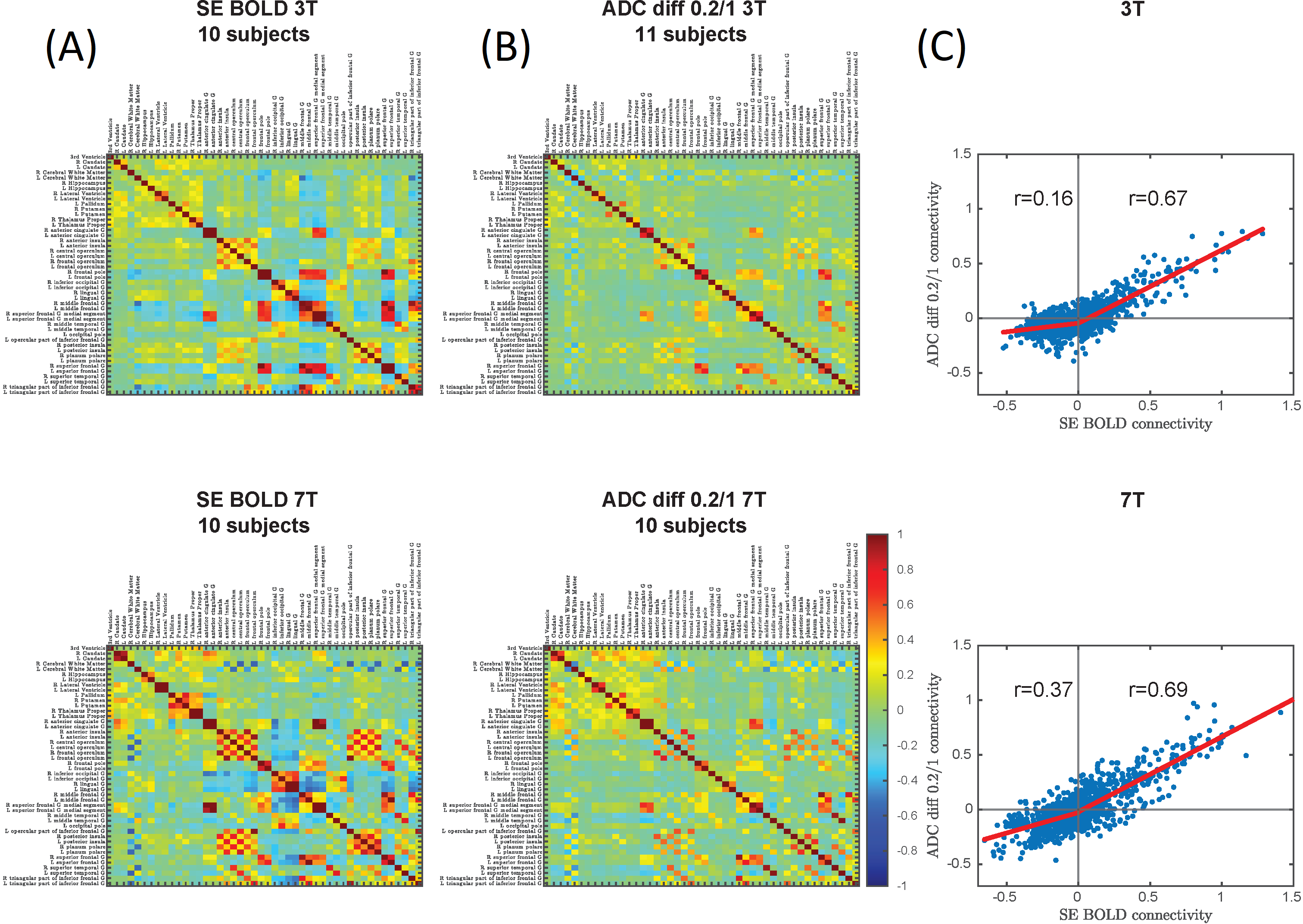
Fig 4: Group averages of Fisher-transformed FC
matrices from SE-BOLD (A) and b=0.2/1 ADC (B) at 3T and 7T. (C) Correlation of
FC strength derived from SE-BOLD vs dfMRI. Positive correlations were somewhat
less pronounced in dfMRI compared to BOLD (r ~ 0.7 for both 3T and 7T), but
anti-correlations were attenuated preferentially in dfMRI, particularly at 3T
(r=0.16).