Luca Vizioli1,2, Steen Moeller1, logan T Dowdle1, Mehmet Akcakaya1, Federico De Martino3, Essa Yacoub1, and Kamil Ugurbil1
1CMRR, University of Minnesota, minneapolis, MN, United States, 2Department of Neurosurgery, University Of Minnesota, minneapolis, MN, United States, 3University Of Maastricht, Maastricht, Netherlands
1CMRR, University of Minnesota, minneapolis, MN, United States, 2Department of Neurosurgery, University Of Minnesota, minneapolis, MN, United States, 3University Of Maastricht, Maastricht, Netherlands
Functional neuroimaging data is often dominated by thermal noise. Using the NORDIC method, which suppresses this noise source, we find substantial signal to noise gains without loss in spatial precision. Activation in one denoised run is comparable to averaging 3 to 5 runs of standard data.
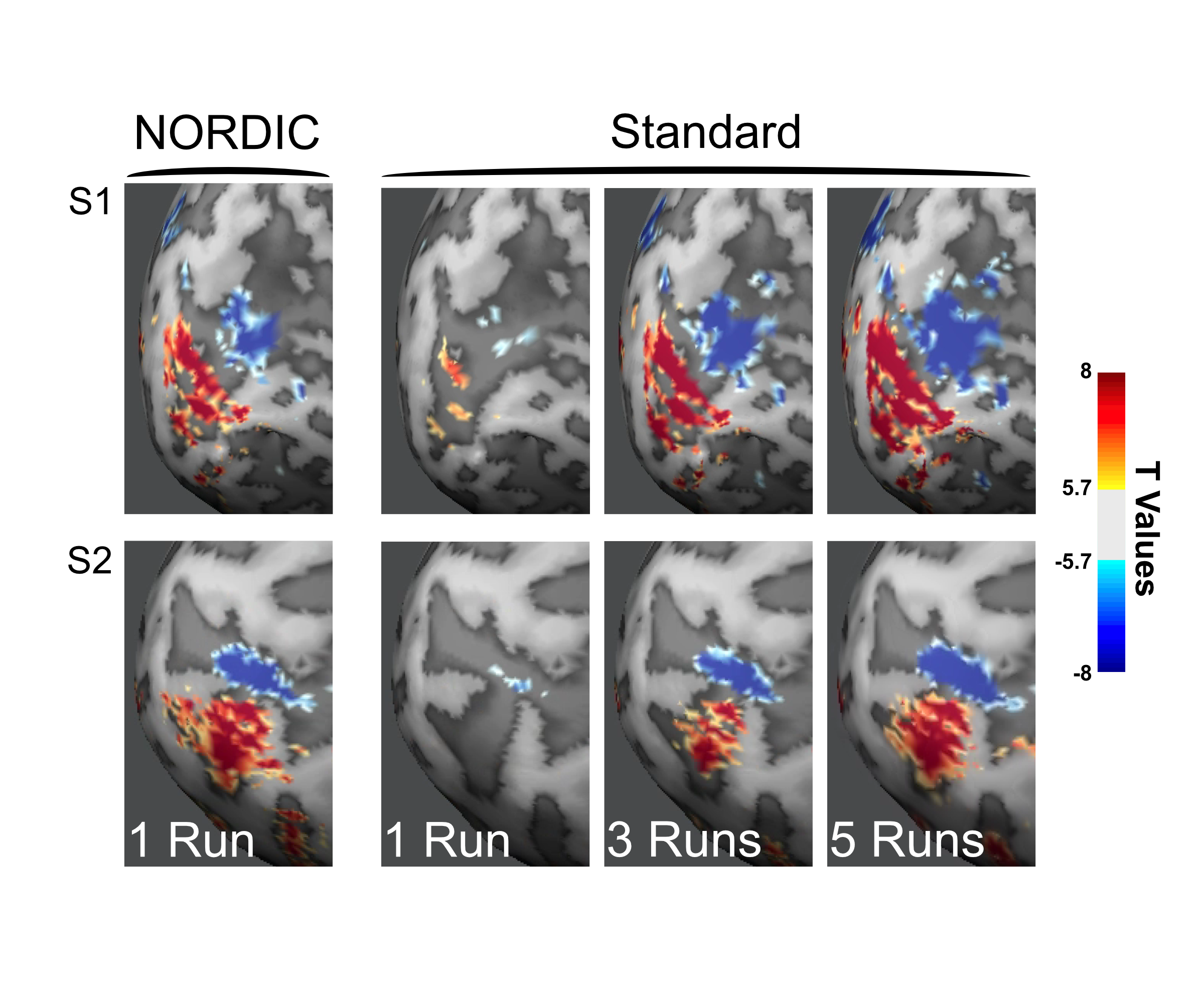
Functional images as t-maps (target > surround) thresholded at t ≥ 5.7 for a single NORDIC processed run (left most panel) and for 1, 3 and 5 Standard processed runs combined, for subject 1 (S1, top row) and subject 2 (S2, lower row). The data were with a 2D GE 0.8 mm iso voxels (iPAT 3; MB2; TR 1350) using a 12 seconds on and 12 seconds off block design where 2 flickering checkerboard (target and surround) were presented 3 times each per run.
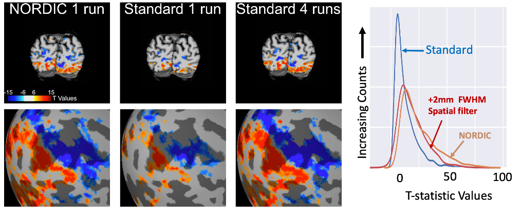
Figure 4: NORDIC denoising on 3T fMRI using the 3T HCP protocol. T-maps with t value ≥ for the contrast target (red) > surround (blue) for a representative slice in the visual cortex (top), and on inflated cortex (bottom). Approximately four runs with standard reconstruction (~10 min of data) are required to achieve the extent of activation comparable a single NORDIC run (~ 2.5 min of data). The right-most panel shows the t-value distribution for NORDIC, Standard, and Standard reconstructions with 2 mm FWHM spatial smoothing, within a the retinotopic representation of the target in V1.