Xingfeng Shao1, Fanhua Guo2, Qinyang Shou1, Kai Wang1, Lirong Yan1,3, Kay Jann1,3, Peng Zhang2, and Danny JJ Wang1,3
1Laboratory of FMRI Technology (LOFT), Mark & Mary Stevens Neuroimaging and Informatics Institute, Keck School of Medicine, University of Southern California, Los Angeles, CA, United States, 2State Key Laboratory of Brain and Cognitive Science, Beijing MRI Center for Brain Research, Institute of Biophysics, Chinese Academy of Sciences, Beijing, China, 3Department of Neurology, University of Southern California, Los Angeles, CA, United States
1Laboratory of FMRI Technology (LOFT), Mark & Mary Stevens Neuroimaging and Informatics Institute, Keck School of Medicine, University of Southern California, Los Angeles, CA, United States, 2State Key Laboratory of Brain and Cognitive Science, Beijing MRI Center for Brain Research, Institute of Biophysics, Chinese Academy of Sciences, Beijing, China, 3Department of Neurology, University of Southern California, Los Angeles, CA, United States
Finger tapping (FT)-induced CBF increase shows a clear ‘double-peak’ pattern (somatosensory input in the superficial layers and motor output in the deep layers). Finger brushing (FB)-induced CBF increase was overall smaller, and mainly peaked in superficial layers (somatosensory input).

Figure 4. (a) CBF map at rest (top) and two activation tasks (FT, middle; FB, bottom). (b) CBF increase evoked by the FT and FB tasks are highlighted in two insets. (c) Laminar profile of CBF at three tasks. (d) Laminar profile of CBF increase evoked by FT and FB. FT induced CBF increase shows a ‘double-peak’ pattern which corresponds to sensory input (superficial) and motor output (deep) layers respectively. FT induced CBF increase shows a weaker increase in superficial layers which corresponds to exteroception sensory input and minimal motor output.
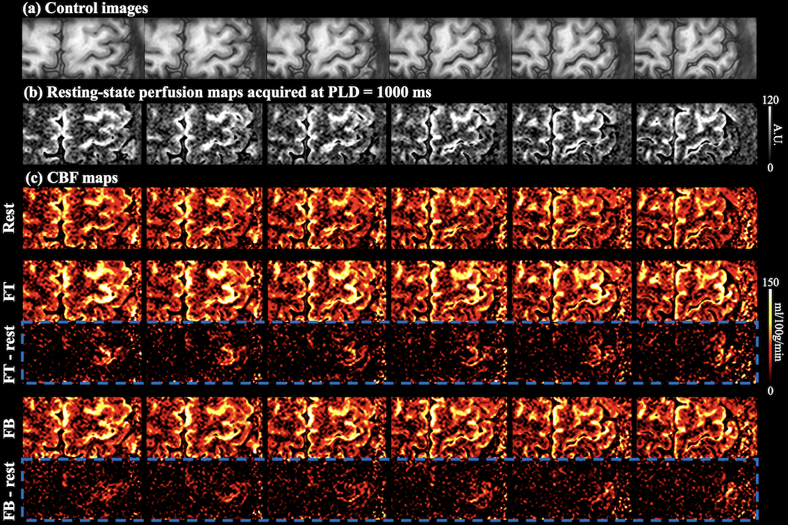
Figure 5. Six axial slices of the control images (a), resting-state perfusion maps (PLD=1000ms) (b) and CBF maps at rest, two activation tasks and the difference between rest and activation (blue boxes) (c). The proposed technique has sufficient coverage for perfusion measurement in M1 and supplementary motor area (SMA), etc.