Wenbo Li1,2, Peter van Zijl1,2, and Qin Qin1,2
1Radiology Department, Johns Hopkins University School of Medicine, Baltimore, MD, United States, 2Kirby Image Center, Kennedy Krieger Institute, Baltimore, MD, United States
1Radiology Department, Johns Hopkins University School of Medicine, Baltimore, MD, United States, 2Kirby Image Center, Kennedy Krieger Institute, Baltimore, MD, United States
A T2-oximetry technique for mapping venous oxygenation based on Fourier-transform based
velocity-selective pulse trains is proposed with the advantage of higher SNR. Preliminary
data on two healthy subjects show that the averaged Yv across all
the voxels is comparable to the global Yv.
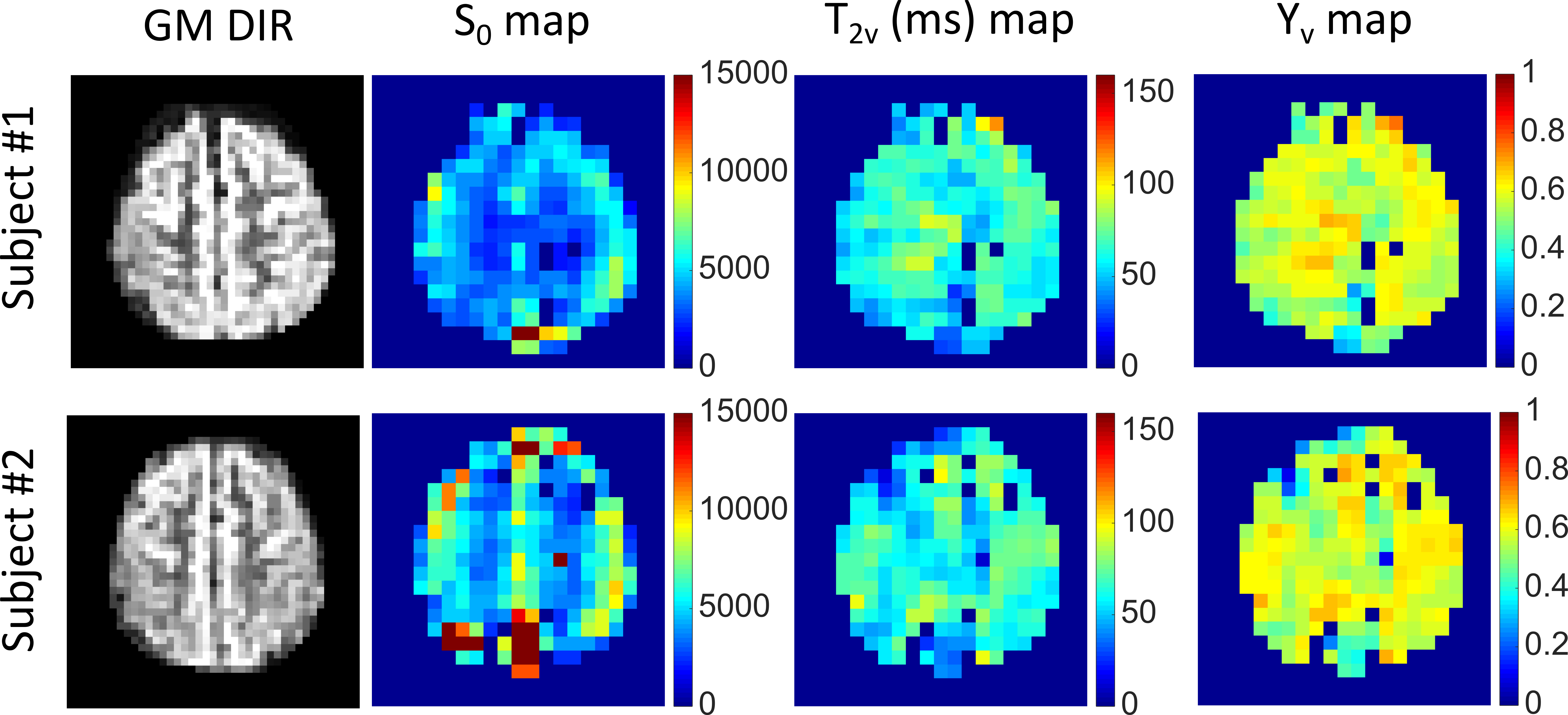
Figure 3. The DIR (GM) map, S0 map, T2v
(ms) map and Yv maps for the two subjects. The extrapolated S0 values are proportional to
the venous CBV and, as expected, show higher signal in the GM than in the WM. The
fitted T2 maps and the corresponding Yv maps reveal a
more uniform contrast between GM and WM, as expected for brain OEF. Several
voxels (mainly in white matter region) with very low signal intensities were excluded
during the data processing.
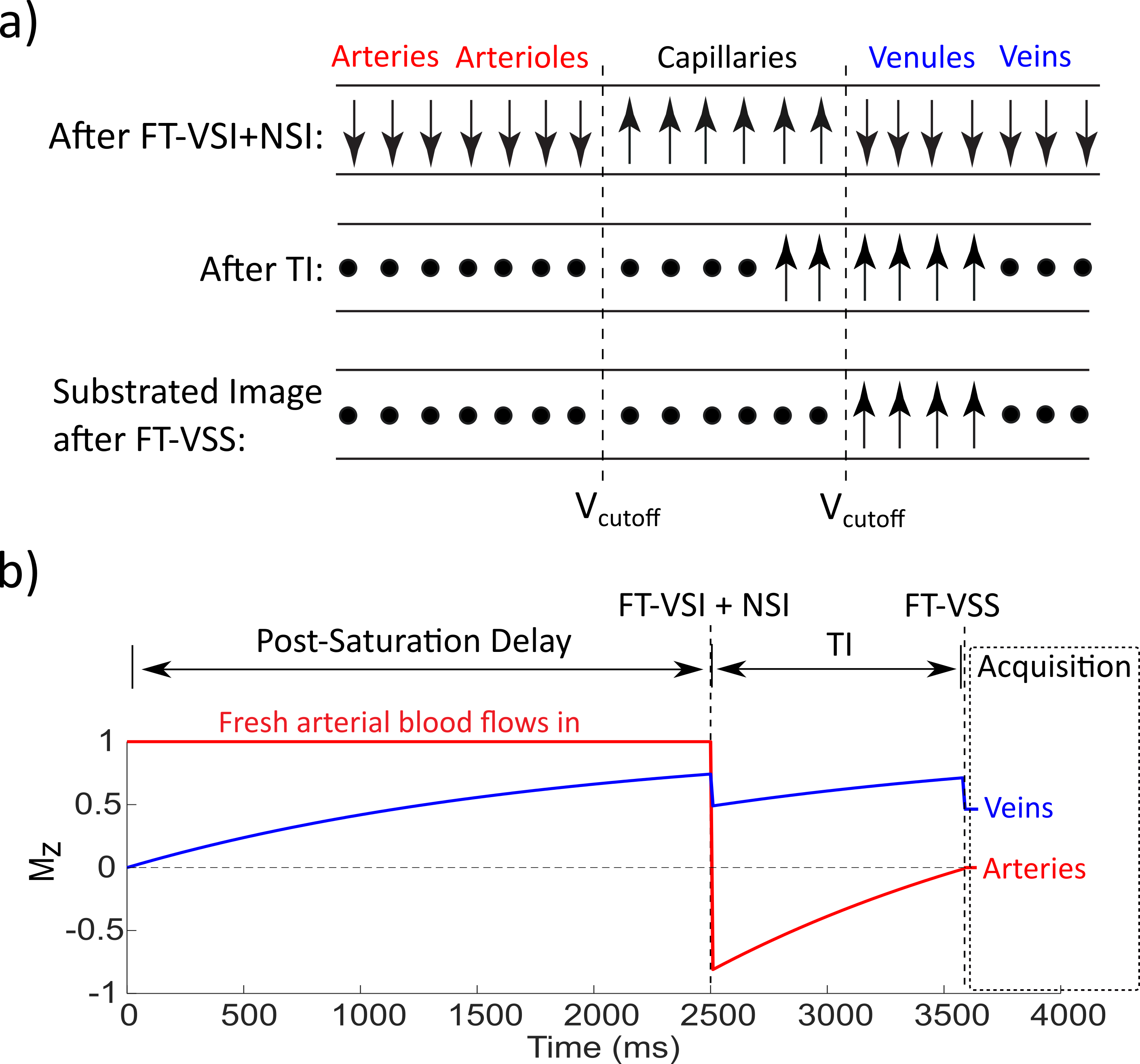
Figure 2.
a) Idealized cartoon depicting the evolution of the longitudinal magnetization
of water in different compartments. Relaxed
and dephased spins are denoted by upright arrows and solid circles,
respectively. b) Simulated magnetization evolution for the arterial and
venous blood. The consecutive FT-VSI and NSI pulses invert spins
flowing above the VCUTOFF for arterial nulling and preserve the spins
moving below the VCUTOFF. These preserved spins will largely outflow
into the venules during TI, thus yielding a high SNR.