Joseph G Woods1, Eric C Wong1, Emma Boyd2, and Divya S Bolar1
1Radiology, UCSD, La Jolla, CA, United States, 2Neurosciences, UCSD, La Jolla, CA, United States
1Radiology, UCSD, La Jolla, CA, United States, 2Neurosciences, UCSD, La Jolla, CA, United States
VESPA ASL is introduced, which simultaneously acquires VSASL and PCASL data. A signal model to quantify cerebral blood flow and arterial transit times (ATT) from these data is presented, with generation of quantitative CBF and ATT maps in vivo.
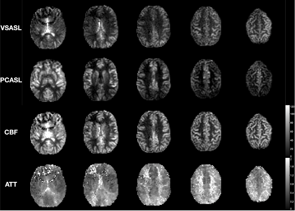
Figure 3: The VESPA decoded VSASL and PCASL data, with the CBF and ATT maps calculated by voxelwise fitting the VESPA signal model to the decoded VESPA data. Five slices from 1 subject are shown. Longer ATTs can be seen in the white matter, posterior circulation, and at both internal and external border zones. Before fitting, the data were voxelwise calibrated by the M0 image and corrected for slice-dependent T1 decay.
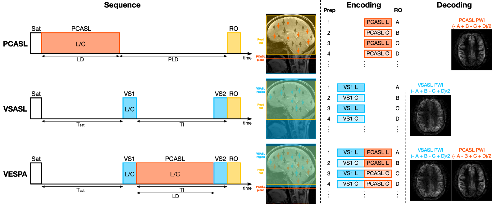
Figure 1: Sequence diagrams, label/control encoding, and contrast decoding strategies for PCASL, VSASL and VESPA. In VESPA, VSASL and PCASL are combined and used to label different pools of inflowing arterial blood. VSASL labeling (VS1) uses a spatially-selective VSASL module surrounding the imaging slab. The PCASL labeling plane is immediately inferior to the VSASL slab. By alternating label and control conditions in different orders, VSASL and PCASL contrasts can be linearly encoded into the acquired images and decoded by simple image arithmetic