Minhui Ouyang1, John Detre2, Chenying Zhao1,3, Samantha Lam1, J. Christopher Edgar1,2, and Hao Huang1,2
1Department of Radiology, The Children's Hospital of Philadelphia, Philadelphia, PA, United States, 2Department of Radiology, Perelman School of Medicine, University of Pennsylvania, Philadelphia, PA, United States, 3Department of Bioengineering, School of Engineering and Applied Science, University of Pennsylvania, Philadelphia, PA, United States
1Department of Radiology, The Children's Hospital of Philadelphia, Philadelphia, PA, United States, 2Department of Radiology, Perelman School of Medicine, University of Pennsylvania, Philadelphia, PA, United States, 3Department of Bioengineering, School of Engineering and Applied Science, University of Pennsylvania, Philadelphia, PA, United States
Heterogenous maturational pattern of regional cerebral
blood flow in infants aged 0-18months, with maturation most prominent in
heteromodal association cortex, was revealed by using a high-resolution 3D
multi-shot, stack-of-spirals pCASL perfusion MRI at isotropic 2.5mm.
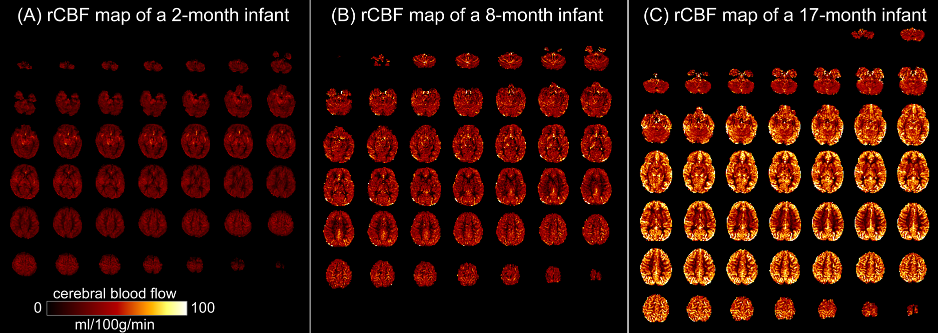
Fig. 2: High-resolution
(2.5x2.5x2.5mm3) regional cerebral blood flow (rCBF) maps acquired
with 3D multi-shot, stack-of-spirals pCASL, from three representative infants
aged 2months (A), 8months (B) and 17months (C).
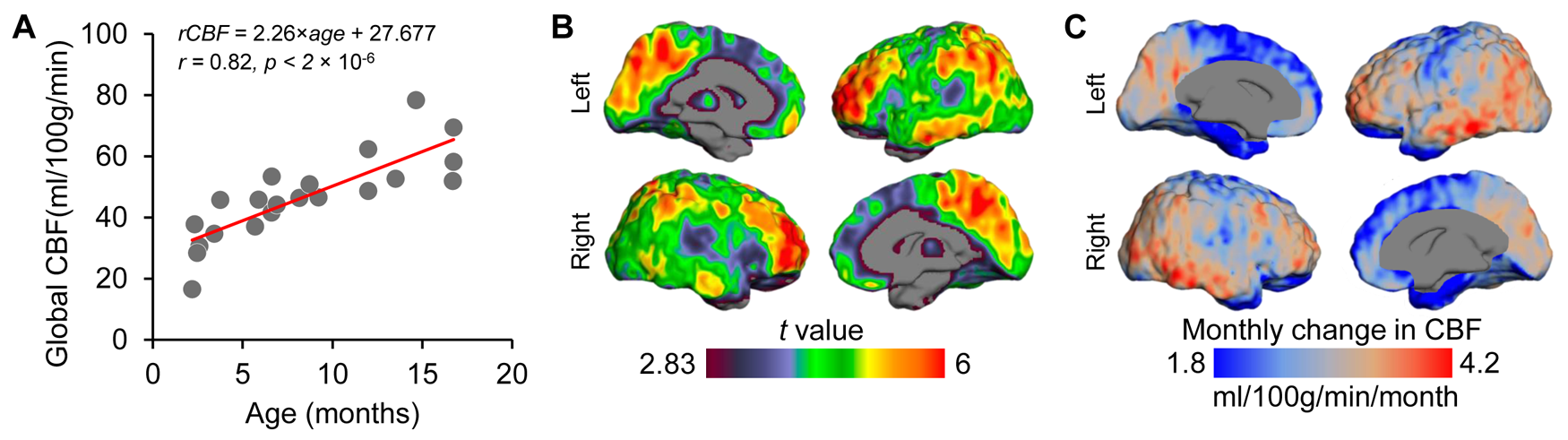
Fig.
3: Main effect of age on cerebral blood flow
(CBF) during infancy. (A) Global CBF increases during infancy. Data points
represent global CBF measured with phase-contrast MRI of each subject. (B) rCBF
increases throughout the cortex, but more prominently in the heteromodal
association cortex. Images thresholded at t(df=21) > 2.83 with p<0.01. (C)
heterogeneous developmental rate of rCBF (ml/100g/min/month) across
cortex during infancy. Slower and faster rCBF growth rates are shown in cool
and warm colors, respectively.