Hualu Han1,2, Zixuan Lin2,3, Melissa Rundle4, Anja Soldan5, Corinne Pettigrew5, Joshua F. Betz6, Kumiko Oishi7, Yang Li2, Binu P. Thomas8, Peiying Liu2, Marilyn Albert5, Denise Park4, and Hanzhang Lu2,3,9
1Center for Biomedical Imaging Research, Department of Biomedical Engineering, School of Medicine, Tsinghua University, Beijing, China, 2The Russell H. Morgan Department of Radiology & Radiological Science, Johns Hopkins University School of Medicine, Baltimore, MD, United States, 3Department of Biomedical Engineering, Johns Hopkins University School of Medicine, Baltimore, MD, United States, 4Center for Vital Longevity, School of Behavioral and Brain Sciences, University of Texas at Dallas, Dallas, TX, United States, 5Department of Neurology, Johns Hopkins University School of Medicine, Baltimore, MD, United States, 6Department of Biostatistics, Johns Hopkins Bloomberg School of Public Health, Baltimore, MD, United States, 7Center for Imaging Science, Johns Hopkins University, Whiting School of Engineering, Baltimore, MD, United States, 8Advanced Imaging Research Center, University of Texas Southwestern Medical Center, Dallas, TX, United States, 9F.M. Kirby Center for Functional Brain Imaging, Kennedy Krieger Institute, Baltimore, MD, United States
1Center for Biomedical Imaging Research, Department of Biomedical Engineering, School of Medicine, Tsinghua University, Beijing, China, 2The Russell H. Morgan Department of Radiology & Radiological Science, Johns Hopkins University School of Medicine, Baltimore, MD, United States, 3Department of Biomedical Engineering, Johns Hopkins University School of Medicine, Baltimore, MD, United States, 4Center for Vital Longevity, School of Behavioral and Brain Sciences, University of Texas at Dallas, Dallas, TX, United States, 5Department of Neurology, Johns Hopkins University School of Medicine, Baltimore, MD, United States, 6Department of Biostatistics, Johns Hopkins Bloomberg School of Public Health, Baltimore, MD, United States, 7Center for Imaging Science, Johns Hopkins University, Whiting School of Engineering, Baltimore, MD, United States, 8Advanced Imaging Research Center, University of Texas Southwestern Medical Center, Dallas, TX, United States, 9F.M. Kirby Center for Functional Brain Imaging, Kennedy Krieger Institute, Baltimore, MD, United States
Longitudinal CBF decrease with age and the longitudinal rate of decline was faster than that from the cross-sectional data. CBF reduction is faster in younger than in older individuals and has spatial differences and hemispheric asymmetry.
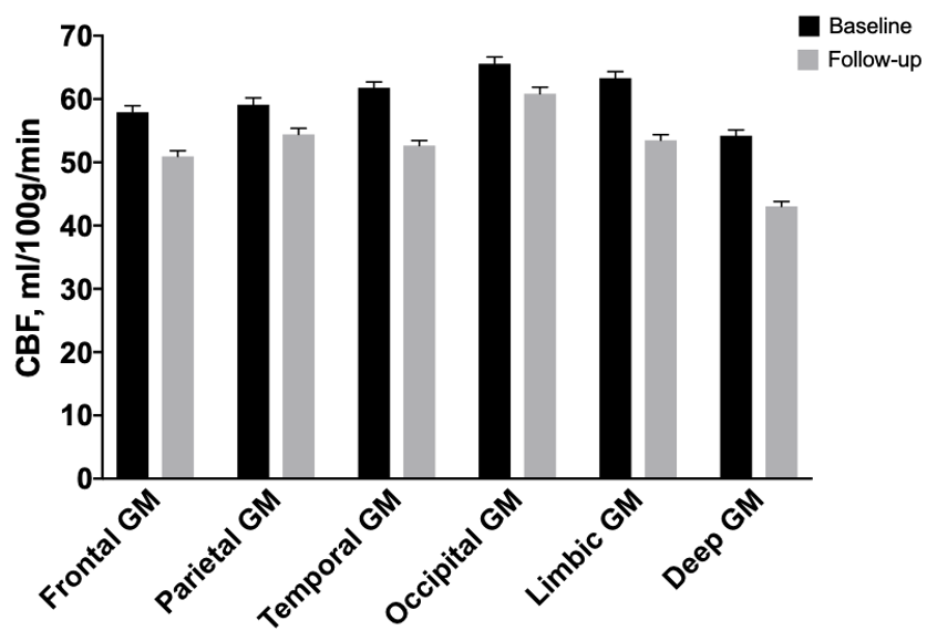
Figure 1. Regional CBF (mean ± standard error) between baseline and follow-up in major GM structures of Study 2.
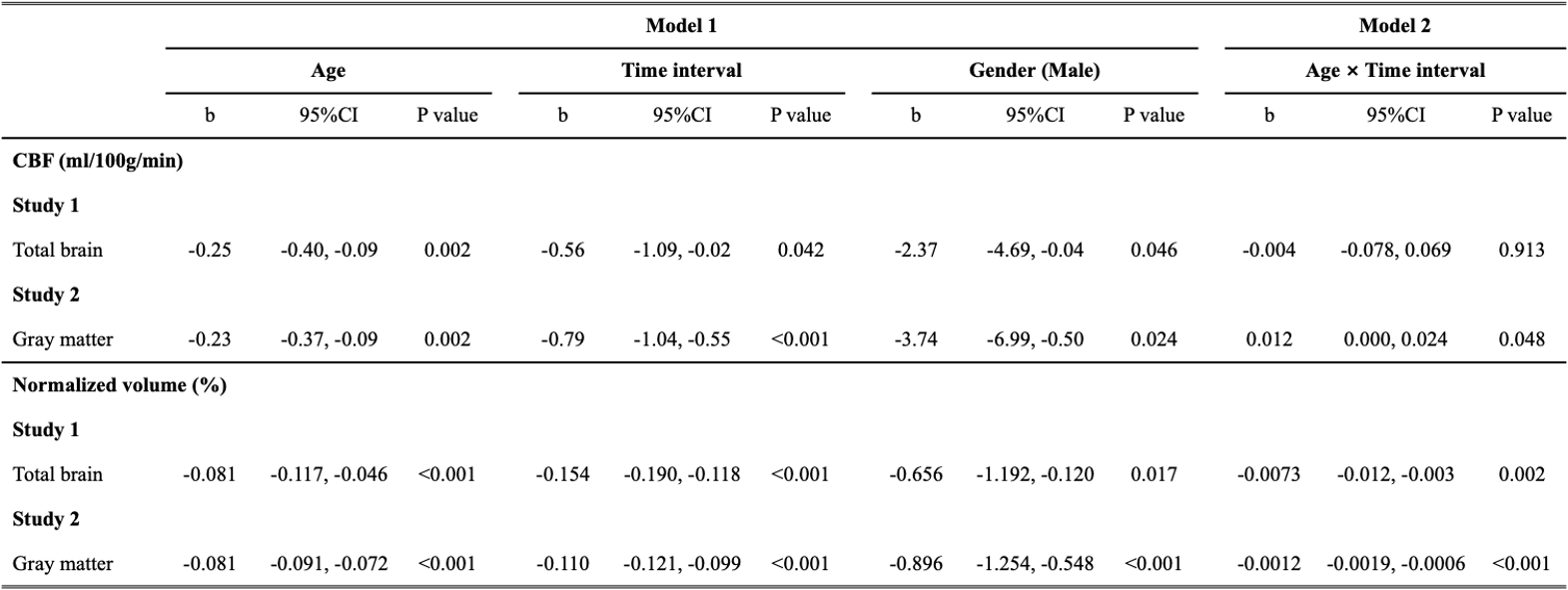
Table 3. Linear mixed models for assessing the changes of global CBF and normalized volume in Study 1 (N=127) and Study 2 (N=115). Age, time interval, gender, GM probability and WM probability were added as the fixed effects in Model 1, and age-by-follow-up time interaction was additionally added in Model 2.