Riccardo Galbusera1,2,3, Erik Bahn4, Matthias Weigel2,3,5, Po-Jui Lu1,2,3, Muhamed Barakovic1,2,3, Reza Rahmanzadeh1,2,3, Peter Dechent6, Antoine Lutti7, Govind Bhagavatheeshwaran8, Ludwig Kappos2,3, Wolfgang Brück4, Christine Stadelmann-Nessler4, and Cristina Granziera1,2,3
1Neurology Clinic and Policlinic, Departments of Medicine, Clinical Research and Biomedical Engineering, University Hospital Basel and University of Basel, Basel, Switzerland, Basel, Switzerland, 2Translational Imaging in Neurology (ThINk) Basel, Department of Biomedical Engineering, University Hospital Basel and University of Basel, Basel, Switzerland, Basel, Switzerland, 3Research Center for Clinical Neuroimmunology and Neuroscience (RC2NB) Basel, University Hospital Basel and University of Basel, Basel, Switzerland, Basel, Switzerland, 4Institute of Neuropathology, University Medical Center, Göttingen, Germany, Göttingen, Germany, 5Division of Radiological Physics, Department of Radiology, University Hospital Basel, Basel, CH, Basel, Switzerland, 6Department of Cognitive Neurology, MR-Research in Neurosciences, University Medical Center Göttingen, Göttingen, Germany, Göttingen, Germany, 7Centre for Research in Neuroscience - Department of Clinical Neurosciences, Laboratoire de recherche en neuroimagerie (LREN) University Hospital and University of Lausanne, Lausanne, Switzerland, Lausanne, Switzerland, 8National Institute of Neurological Disorders and Stroke, Bethesda, MD, USA, Bethesda, MD, United States
1Neurology Clinic and Policlinic, Departments of Medicine, Clinical Research and Biomedical Engineering, University Hospital Basel and University of Basel, Basel, Switzerland, Basel, Switzerland, 2Translational Imaging in Neurology (ThINk) Basel, Department of Biomedical Engineering, University Hospital Basel and University of Basel, Basel, Switzerland, Basel, Switzerland, 3Research Center for Clinical Neuroimmunology and Neuroscience (RC2NB) Basel, University Hospital Basel and University of Basel, Basel, Switzerland, Basel, Switzerland, 4Institute of Neuropathology, University Medical Center, Göttingen, Germany, Göttingen, Germany, 5Division of Radiological Physics, Department of Radiology, University Hospital Basel, Basel, CH, Basel, Switzerland, 6Department of Cognitive Neurology, MR-Research in Neurosciences, University Medical Center Göttingen, Göttingen, Germany, Göttingen, Germany, 7Centre for Research in Neuroscience - Department of Clinical Neurosciences, Laboratoire de recherche en neuroimagerie (LREN) University Hospital and University of Lausanne, Lausanne, Switzerland, Lausanne, Switzerland, 8National Institute of Neurological Disorders and Stroke, Bethesda, MD, USA, Bethesda, MD, United States
We have
identified the imaging correlates of multiple sclerosis lesion subtypes by
exploiting post-mortem multiparametric quantitative MRI and histopathological
analysis. Remyelinated lesions showed distinct MRI characteristics compared to
other MS lesions.
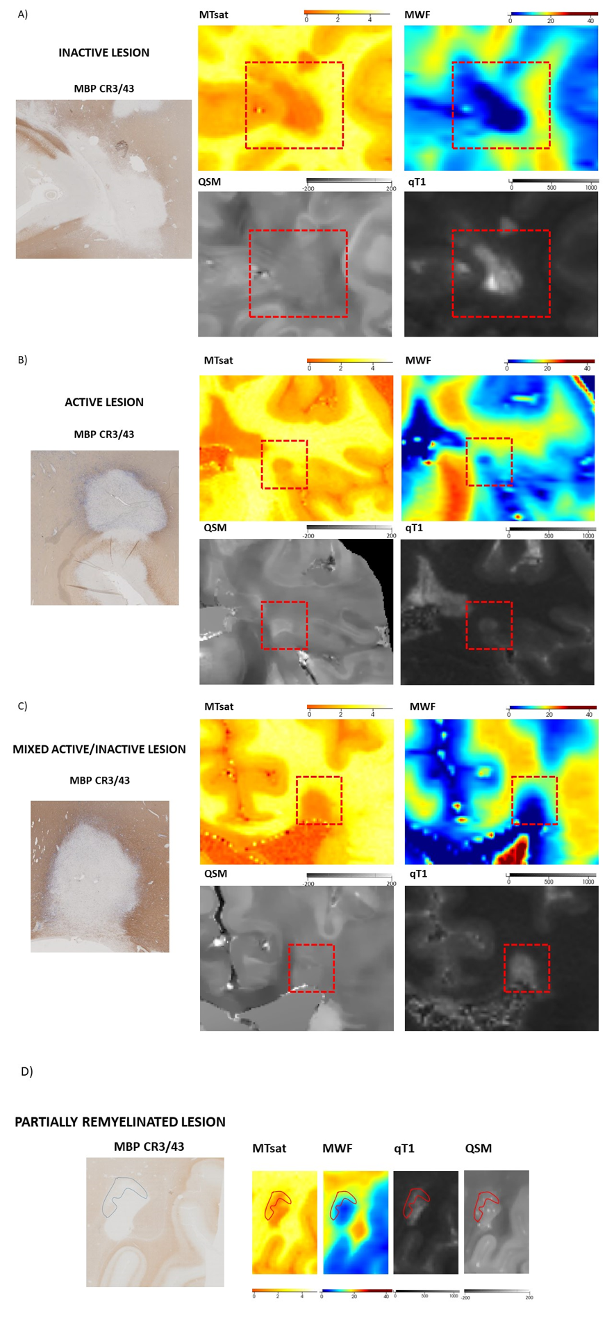
Figure 1. Immunohistological staining for myelin basic
protein (MBP) and for CR3/43 (left) and parametric maps (MTsat, MWF, qT1, QSM) showing
the characteristics of different histopathological MS lesions subtypes in
(right): A) inactive lesion; (B) active lesion; (C) mixed active/inactive lesions and
(D) remyelinated part of a lesion.
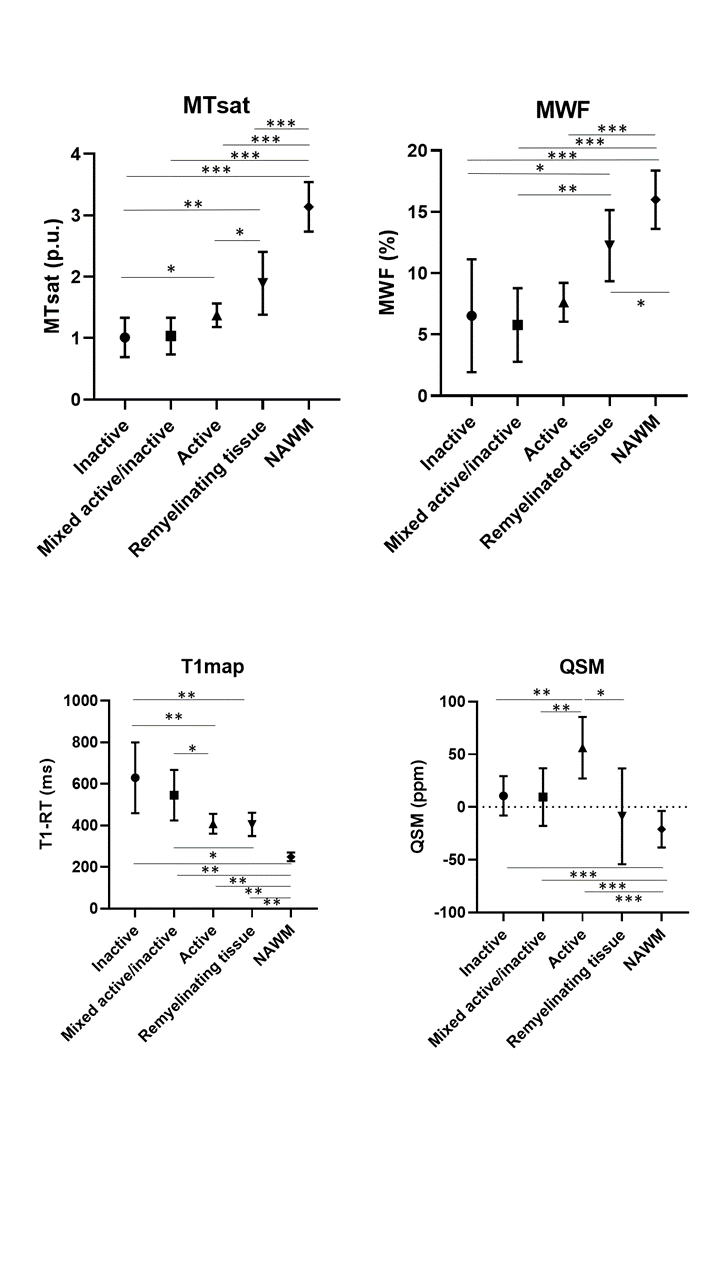
Figure 2. Quantitative
MRI measures across different lesion categories and NAWM. *** p < 0,0001; **
p< 0,001, * p< 0,05.