Jianpan Huang1, Jiadi Xu2,3, Joseph H. C. Lai1, Henry K. F. Mak4, Koon Ho Chan5, and Kannie W. Y. Chan1,3,6
1Department of Biomedical Engineering, City University of Hong Kong, Hong Kong, China, 2F.M. Kirby Research Center for Functional Brain Imaging, Kennedy Krieger Research Institute, Baltimore, MD, United States, 3Russell H. Morgan Department of Radiology and Radiological Science, The Johns Hopkins University School of Medicine, Baltimore, MD, United States, 4Department of Diagnostic Radiology, Li Ka Shing Faculty of Medicine, The University of Hong Kong, Hong Kong, China, 5Department of Medicine, Li Ka Shing Faculty of Medicine, The University of Hong Kong, Hong Kong, China, 6City University of Hong Kong Shenzhen Research Institute, Shenzhen, China
1Department of Biomedical Engineering, City University of Hong Kong, Hong Kong, China, 2F.M. Kirby Research Center for Functional Brain Imaging, Kennedy Krieger Research Institute, Baltimore, MD, United States, 3Russell H. Morgan Department of Radiology and Radiological Science, The Johns Hopkins University School of Medicine, Baltimore, MD, United States, 4Department of Diagnostic Radiology, Li Ka Shing Faculty of Medicine, The University of Hong Kong, Hong Kong, China, 5Department of Medicine, Li Ka Shing Faculty of Medicine, The University of Hong Kong, Hong Kong, China, 6City University of Hong Kong Shenzhen Research Institute, Shenzhen, China
A pulsed-CEST imaging scheme was applied to acquire relayed nuclear Overhauser effect weighted (rNOEw) images of human brain at 3T and significant lower rNOEw signal were found in multiple sclerosis (MS) brains compared to neuromyelitis optica (NMO) and healthy brains.
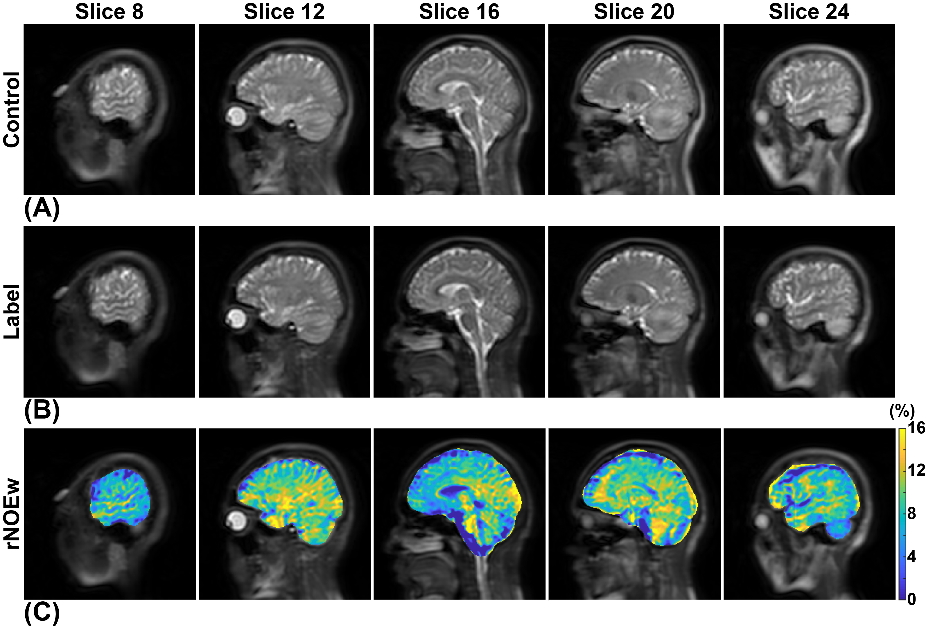
FIGURE 1. An exemplary illustration of generating rNOE weighted (rNOEw) images (C) using control images (A) and label images (B).
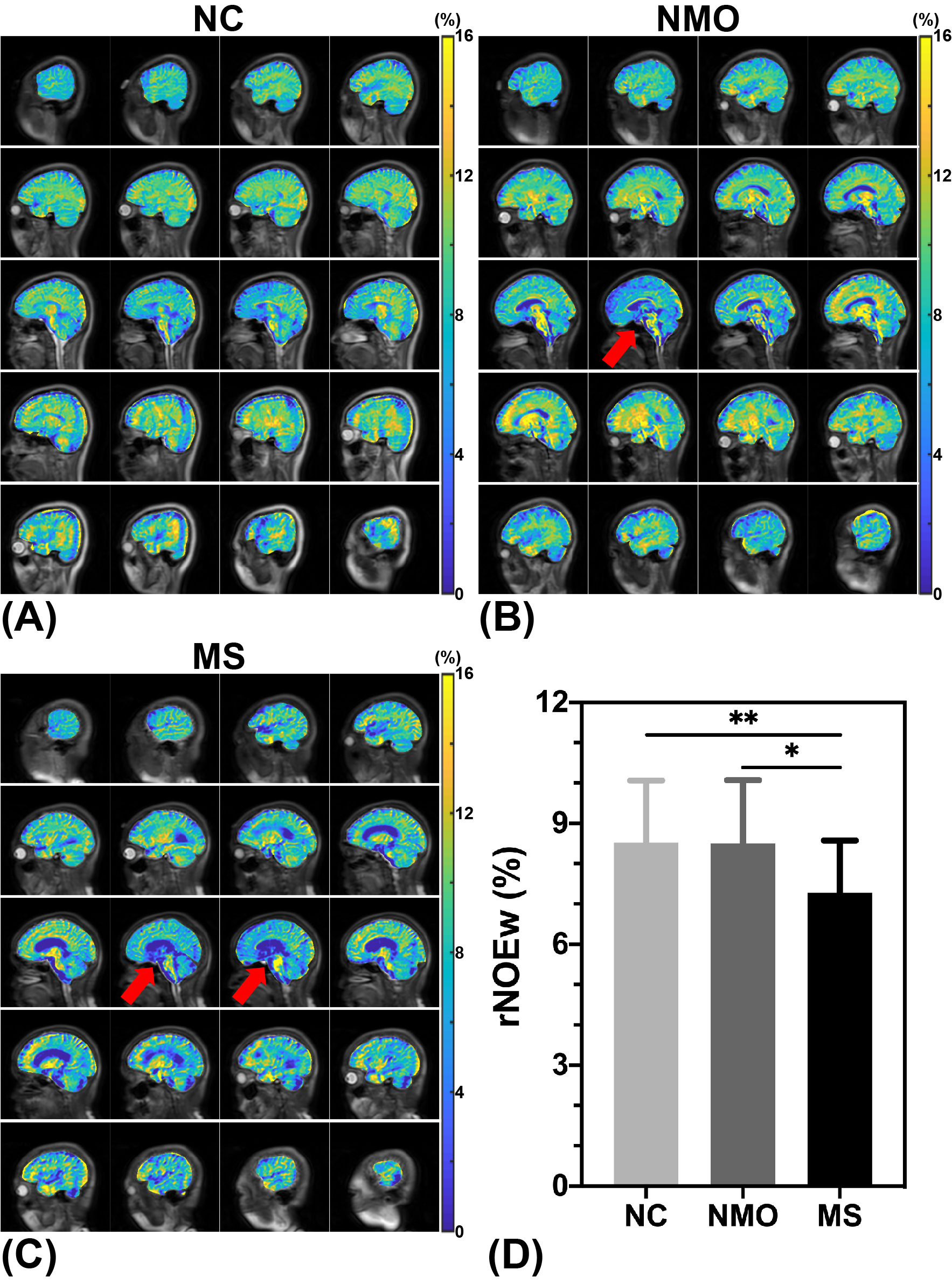
FIGURE 2. Representative rNOEw images of NC (A), NMO (B) and MS (C). (D) comparison of rNOEw signal among three cohorts of subjects (NC = 20, NMO = 15, MS = 20). Significance levels: *, p<0.05; **, p<0.01.