Tianjia Zhu1,2, Qiyuan Tian3,4, Susie Huang3,4, and Hao Huang1,5
1Department of Radiology, Children's Hospital of Philadelphia, Philadelphia, PA, United States, 2Department of Bioengineering, University of Pennsylvania, Philadelphia, PA, United States, 3Department of Radiology, Harvard Medical School, Boston, MA, United States, 4Massachusetts General Hospital, Boston, MA, United States, 5Department of Radiology, University of Pennsylvania, Philadelphia, PA, United States
1Department of Radiology, Children's Hospital of Philadelphia, Philadelphia, PA, United States, 2Department of Bioengineering, University of Pennsylvania, Philadelphia, PA, United States, 3Department of Radiology, Harvard Medical School, Boston, MA, United States, 4Massachusetts General Hospital, Boston, MA, United States, 5Department of Radiology, University of Pennsylvania, Philadelphia, PA, United States
We have demonstrated the sensitivity of
cortical kurtosis measurement to diffusion time formulated in
Kurtosis-based Imaging of Neurite and Soma Architecture (KINSA) modeling using
both with Connectome scanner dMRI and simulation studies.
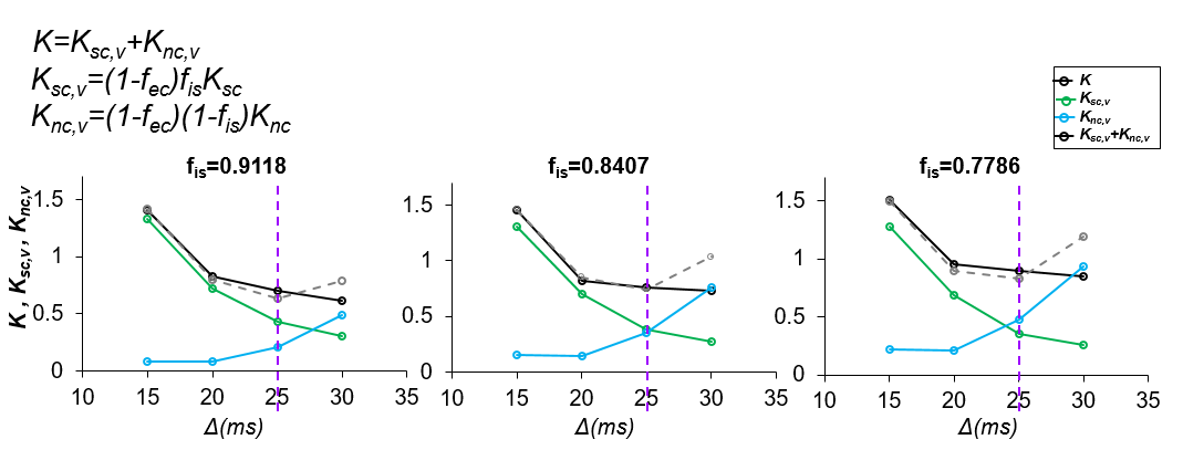
Fig 2. Both somas and neurites
contribute to mean kurtosis in the cerebral cortex and total kurtosis as well
as Ksc,v, Knc,v are sensitive to diffusion time. Both soma Ksc,v=(1-fec)fisKsc, (green lines) and neurite Knc,v=(1-fec)(1-fis)Knc, (blue lines) contribute to total intracellular kurtosis K
across diffusion times and various soma volume fractions. As demonstrated by
the vertical purple dashed lines, Ksc,v+Knc,v=K as
expected for
25ms in
the case fec=0.
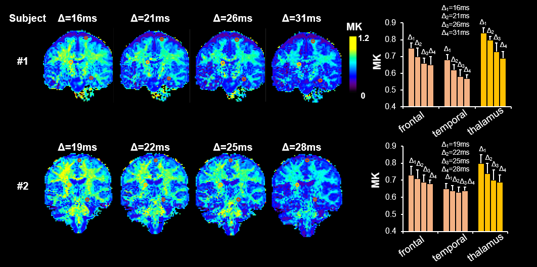
Fig 4. Cortical mean kurtosis(MK) is sensitive
to diffusion time in in-vivo data. Left:
Significant total MK change across relevant diffusion times 16-31ms for both
subjects. Right: Bar plot for MK values in ROIs chosen for the frontal,
temporal, thalamus also quantitatively shown significant MK drop.