Lucas Soustelle1,2, Thomas Troalen3, Andreea Hertanu1,2, Maxime Guye1,2, Jean-Philippe Ranjeva1,2, Guillaume Duhamel1,2, and Olivier M. Girard1,2
1Aix Marseille Univ, CNRS, CRMBM, Marseille, France, 2APHM, Hôpital Universitaire Timone, CEMEREM, Marseille, France, 3Siemens Healthcare SAS, Saint-Denis, France
1Aix Marseille Univ, CNRS, CRMBM, Marseille, France, 2APHM, Hôpital Universitaire Timone, CEMEREM, Marseille, France, 3Siemens Healthcare SAS, Saint-Denis, France
Fast MPF mapping has shown great promises for the evaluation of
myelin-related studies while allowing for acceptable scan times. Here we show how
the input T1 map of the single-point method inherently biases MPF values, and
that T1 should be jointly estimated in SP-quantitative MT applications.
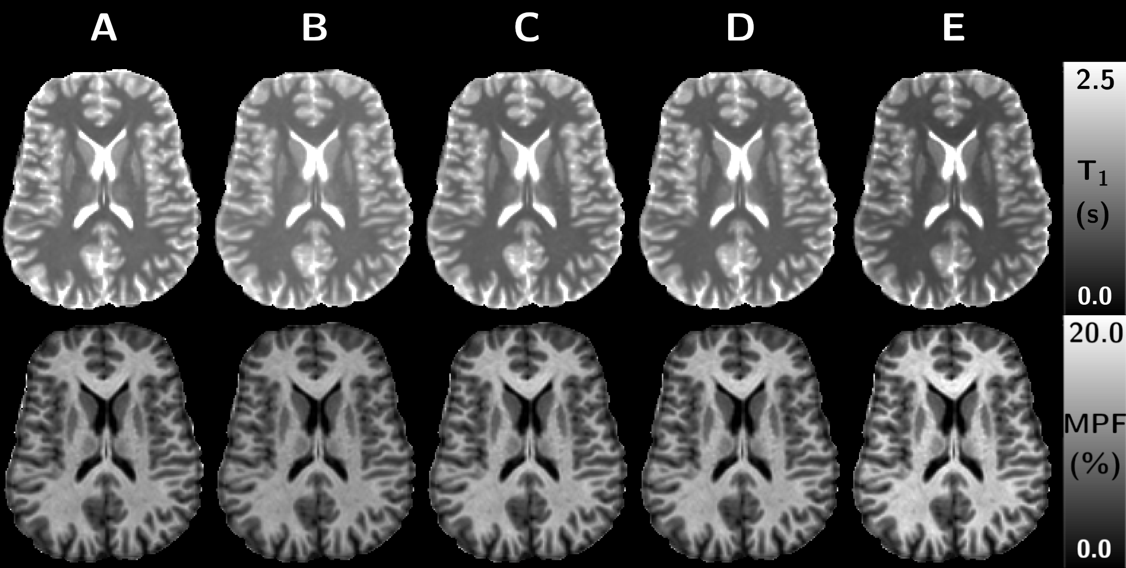
Figure 2: Representative axial slices of T1 (upper row) and MPF (lower row)
maps from VFA protocols A to E.

Figure 1: Simulated difference (ΔT1) between input T1 (1000 ms) and estimated
T1 by the VFA method as a function of the pulse width for an SPGR sequence
without MT effects (orange) or perfect spoiling (yellow), SPGR with MT and
realistic spoiling (blue), and SPGR with CSMT pulses with MT and realistic
spoiling at reference FA of 30° (purple) and 90° (green). Simulated bias related
to protocols A to E are reported. SPGR-CSMT estimations were performed with a
pulse duration superior to 1-ms as spectral overlapping was found suitably
limited to not disrupt the on-resonance component.
