Jelle Veraart1, Erika P. Raven1, Luke J. Edwards2, Nikolaus Weiskopf2,3, and Derek K. Jones4,5
1Center for Biomedical Imaging, NYU Grossman School of Medicine, New York, NY, United States, 2Max Planck Institute for Human Cognitive and Brain Sciences, Leipzig, Germany, 3Felix Bloch Institute for Solid State Physics, Leipzig University, Leipzig, Germany, 4School of Psychology, Cardiff University, Cardiff, United Kingdom, 5Mary MacKillop Institute for Health Research, Australian Catholic University, Melbourne, Australia
1Center for Biomedical Imaging, NYU Grossman School of Medicine, New York, NY, United States, 2Max Planck Institute for Human Cognitive and Brain Sciences, Leipzig, Germany, 3Felix Bloch Institute for Solid State Physics, Leipzig University, Leipzig, Germany, 4School of Psychology, Cardiff University, Cardiff, United Kingdom, 5Mary MacKillop Institute for Health Research, Australian Catholic University, Melbourne, Australia
The non-invasive quantification of the axon diameter in the human white matter using diffusion MRI is feasible, yet limited to the larger axons. The technique yields a reproducible and sensitive metric, with strong inter- and along-tract variability in the human white matter.
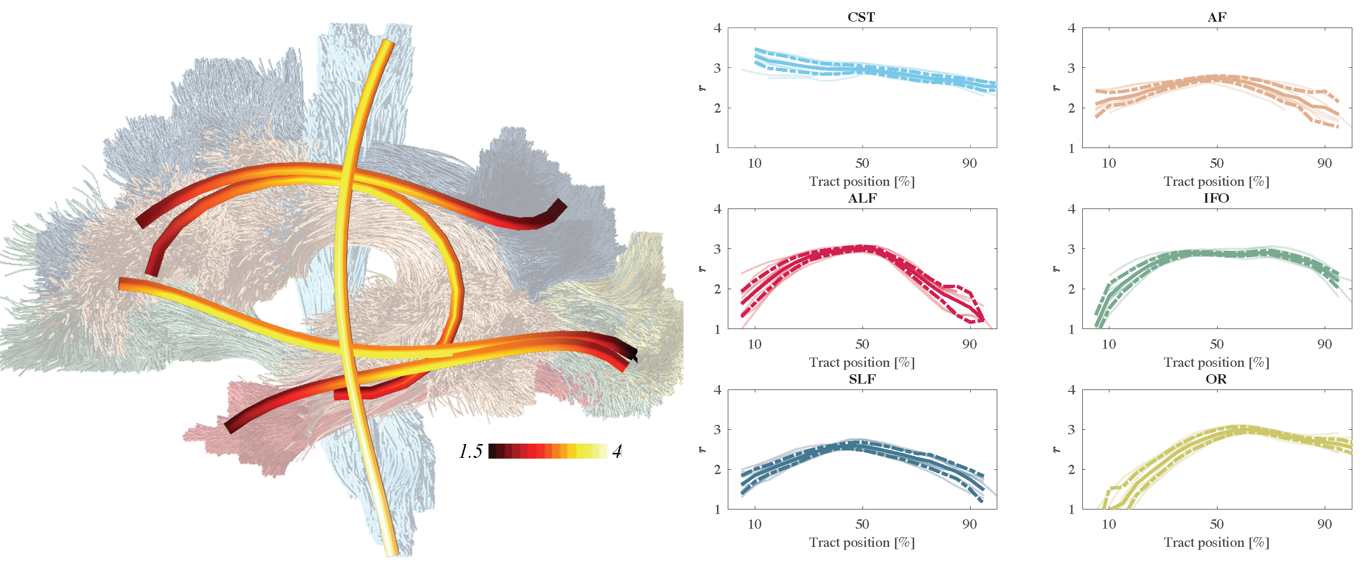
Figure 4: The trend of the effective MR radius r [μm] along the tract (posterior to anterior or inferior to superior) for each individual measurements (5 subjects and 2 repetitions) are shown in shaded lines. In addition, we show the average (solid) and 95% confidence intervals (dashed).
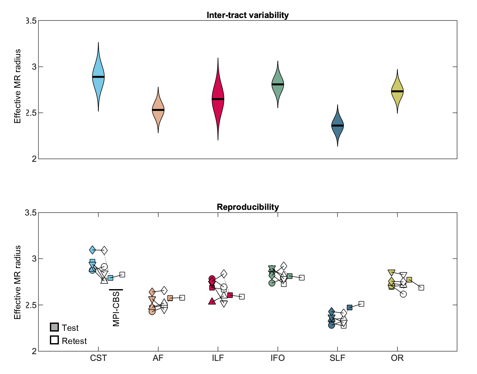
Figure 2: (top) The distribution of MR axon radii [μm]. (bottom) The average MR axon radii for each individual subject (markers) is shown for all scan scan sessions.