Jaakko Paasonen1, Petteri Stenroos1,2, Hanne Laakso1, Tiina Pirttimäki1, Ekaterina Zhurakovskaya1, Raimo A Salo1, Heikki Tanila1, Djaudat Idiyatullin3, Michael Garwood3, Shalom Michaeli3, Silvia Mangia3, and Olli Gröhn1
1A.I.V. Institute for Molecular Sciences, University of Eastern Finland, Kuopio, Finland, 2Grenoble Institut des Neurosciences, Grenoble, France, 3Center for Magnetic Resonance Research, University of Minnesota, Minneapolis, MN, United States
1A.I.V. Institute for Molecular Sciences, University of Eastern Finland, Kuopio, Finland, 2Grenoble Institut des Neurosciences, Grenoble, France, 3Center for Magnetic Resonance Research, University of Minnesota, Minneapolis, MN, United States
Here we introduce a novel approach for fMRI studies in head-fixed and
minimally restrained rats that can express behavior. Our approach links global
network activity to behavior and has potential to enable novel experimental designs
in neuroscience studies.
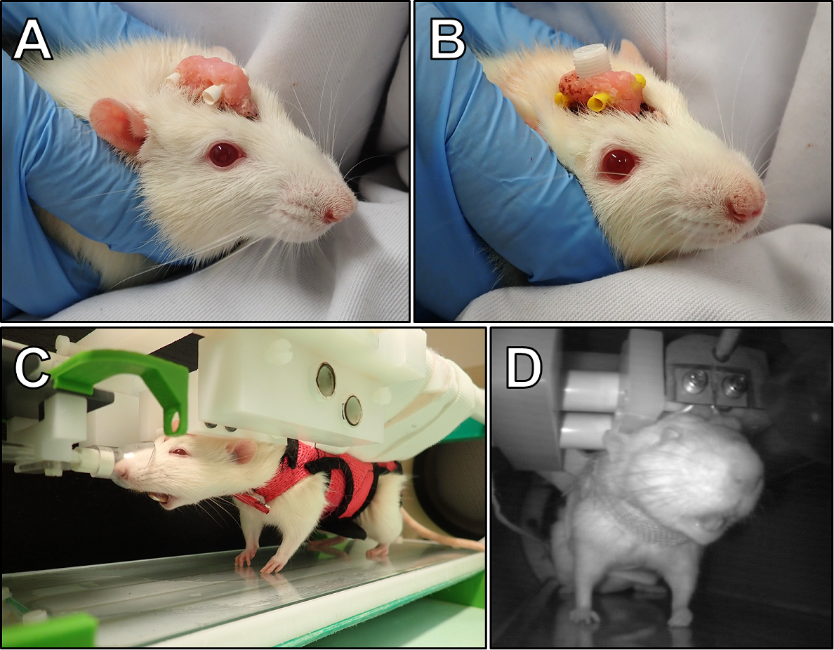
Figure
1. The implant for head-fixation (A) with EEG electrode connector (B), a
head-fixed rat wearing walking harness in a custom-made habituation and imaging
holder for behavioral studies (C), and a habituated rat inside the 9.4T MRI
scanner.
Two 2 mm pins penetrating the implant (A, B) allow a robust head-fixation (C).
Warm-water circulation kept the platform of the holder warm (C). The nose cone was
remotely removed (compare C and D) and returned when necessary. A single loop
transmitter-receiver coil was placed around the implant and below the
head-fixing pins (D) to conduct MRI.
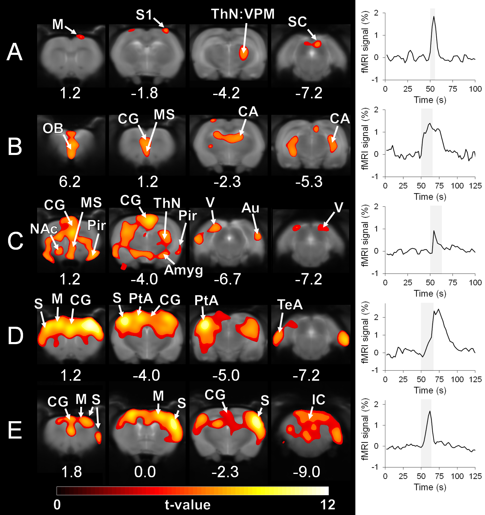
Figure
4. Activation maps (A-E) obtained during spontaneous behavior. Timings of the behavior are shown on the fMRI time
series as shaded regions. The block design analysis included only a single
event. In A, the rat was whisking. In B, the rat was sniffing. In C, the rat expressed
increased arousal. In D, the rat tried to walk. In E, the rat tried to walk but
paws slipped. The maps are overlaid on anatomical images. The slight mismatch between
images and maps originates from the susceptibility-induced signal void in anatomical
images that is not present in the MB-SWIFT images.