1NeuroSpin, Gif-sur-Yvette, France, 2Shionogi & Co., Ltd., Osaka, Japan

Figure 2: Example of results in a single subject
Top: BOLD fMRI showing Superior Colliculus and line position.
Middle: 2D/3D renderings of the space-time activation pattern (b=0) ([9 9] median filtering). The activation spot extends beyond the end of the stimulation.
Bottom: id with b=1800s/mm² showing 2 peaks with slightly different spatial extents, one inside the activation window and the other after the stimulation
Right: Response time courses. The response amplitude (beta) is higher for b=1800s/mm² and present 2 peaks. Both responses are statistically significant.
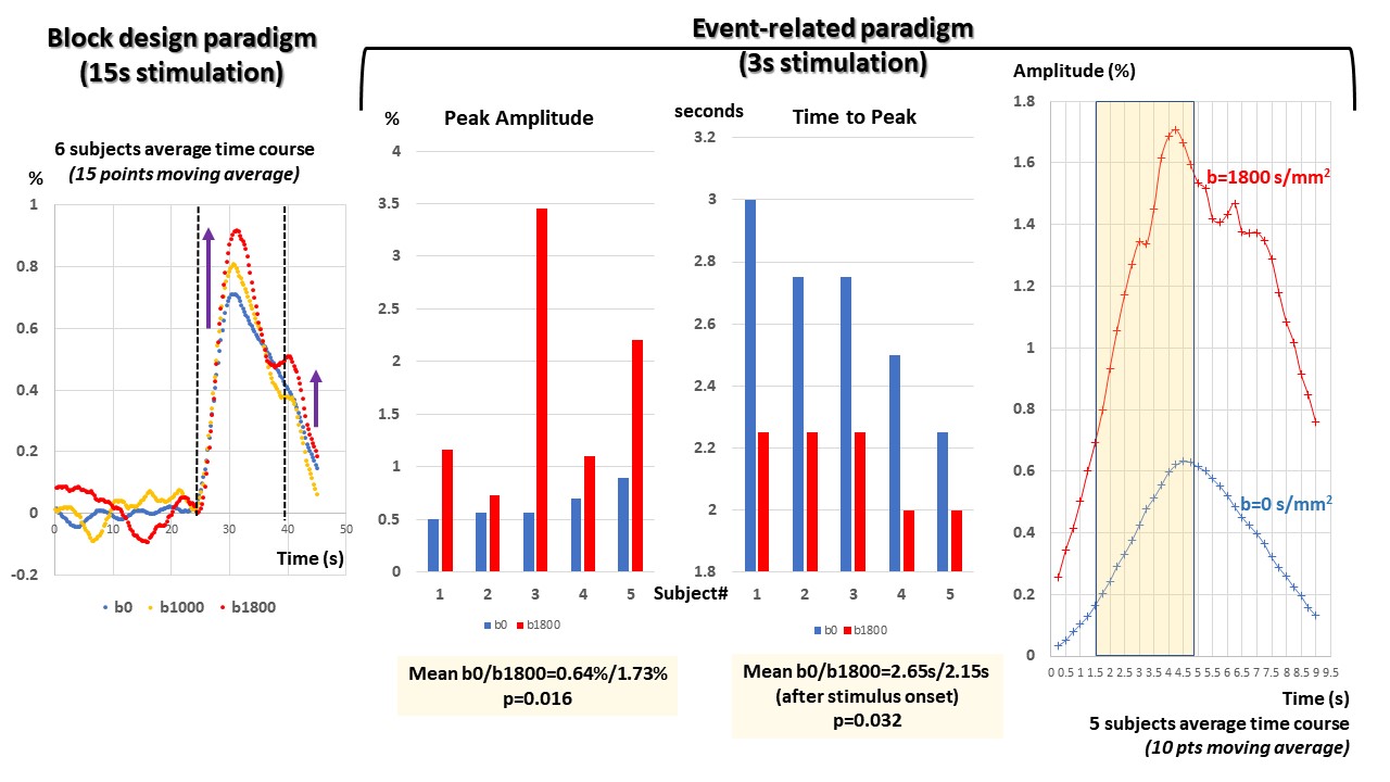
Figure 3: Group results
Left: Group average (n=6) for BP (15 points moving average). The amplitude increase with b value of the main peak and the shoulder are clearly visible.
Middle: Individual results (5 subjects) showing the differences in amplitude and in time-to-peak between b=0 and 1800s/mm² (p=0.016 and p=0.032, respectively).
Right: Group average (n=5) for ER (10 points moving average).