Davide Boido1, Ali-Kémal Aydin2, Yannick Goulam Houssen2, Demené Charlie3, Mickael Tanter3, Serge Charpak2, and Luisa Ciobanu1
1NeuroSpin, CEA, Paris, France, 2Inserm - Institut de la Vision, Paris, France, 3Physics for Medicine, ESPCI, INSERM, CNRS, PSL Research University, Paris, France
1NeuroSpin, CEA, Paris, France, 2Inserm - Institut de la Vision, Paris, France, 3Physics for Medicine, ESPCI, INSERM, CNRS, PSL Research University, Paris, France
With BOLD fMRI at UHF we recorded the mouse olfactory bulb at unprecedented spatio-temporal resolution, detecting sharp responses to 1s odor and multimodal responses at higher odor strengths. With functional ultrasound co-registration we investigated CBV - BOLD relationship in small ROIs.
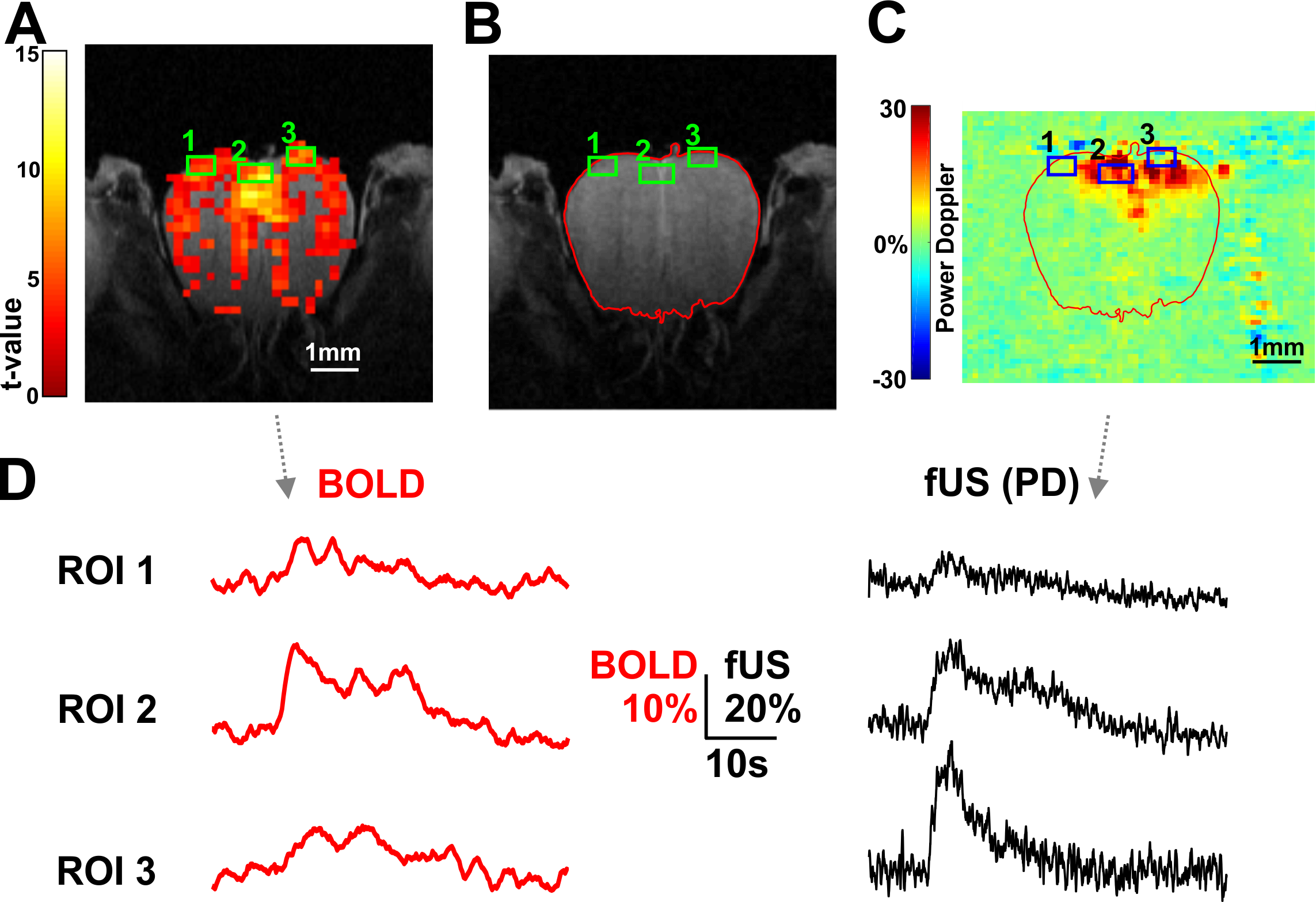
(A) Statistical BOLD activation
map of a coronal section of the OB. ROIs were arbitrary chosen and represented
by green frames. (B) ROIs were reported onto RARE anatomical acquisitions and
(C) transferred to fUS-PD maps by means of the co-registration. (D) BOLD (red)
and fUS-PD (black) time courses within each ROI: the correlation between the 2
signals is apparent.
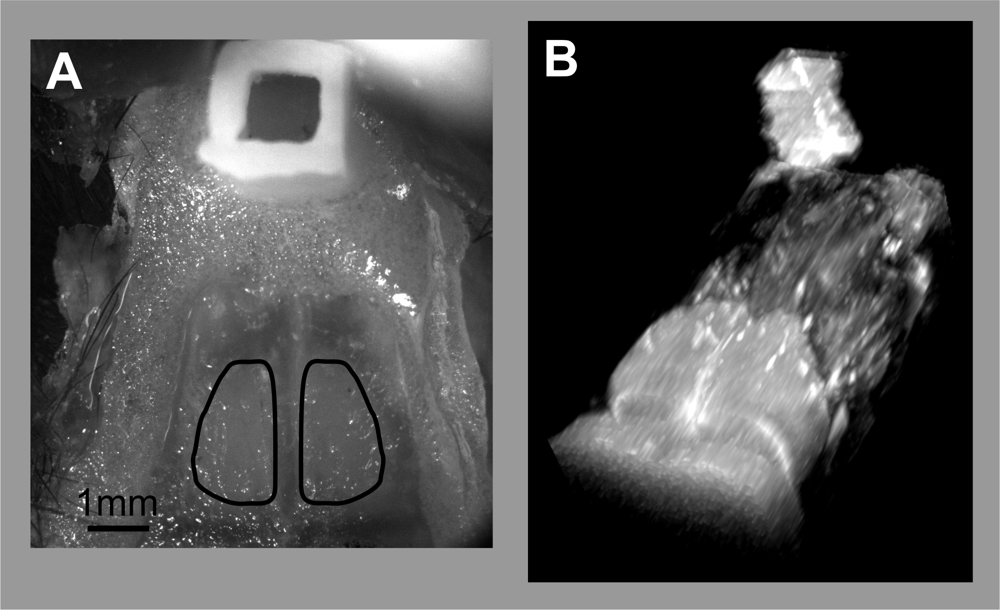
(A) top view of the bone over the
OB and the 3D-printed plastic reservoir. The shape on the OB under the bone is
depicted by the black lines. (B) 3D reconstruction from a RARE acquisition of
the ensemble OB + agar-filled reservoir. Under the reservoir is the nasal
cavity with the fibers connecting the olfactory epithelium to the OB.