1Center for Neuroscience Imaging Research (CNIR), Institute for Basic Science (IBS), Suwon, Korea, Republic of, 2Department of Biomedical Engineering, Sungkyunkwan University, Suwon, Korea, Republic of
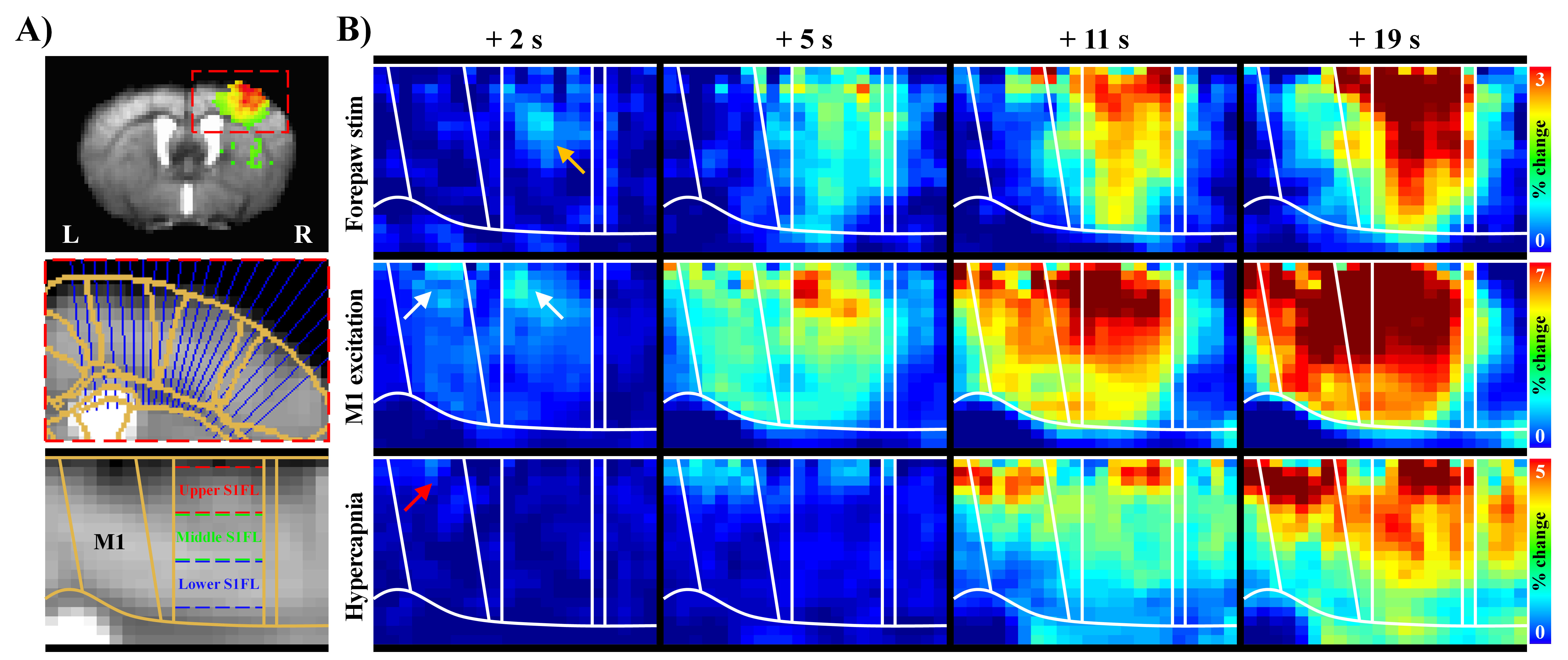
Figure 1. Laminar BOLD responses to neural stimulation and hypercapnic challenge have different origins.
A) The cortical areas (S1FL and M1) were linearized using radially projecting lines perpendicular to the cortical edges.
B) Dynamic BOLD changes were calculated with average window of 2 s duration at interval of 1 s. Sensory-evoked responses first appeared in S1FL middle layer (yellow arrow), whereas early responses by M1 excitation in upper areas of M1 and S1FL (white arrows), and hypercapnic responses started in M1 upper layer (red arrow).
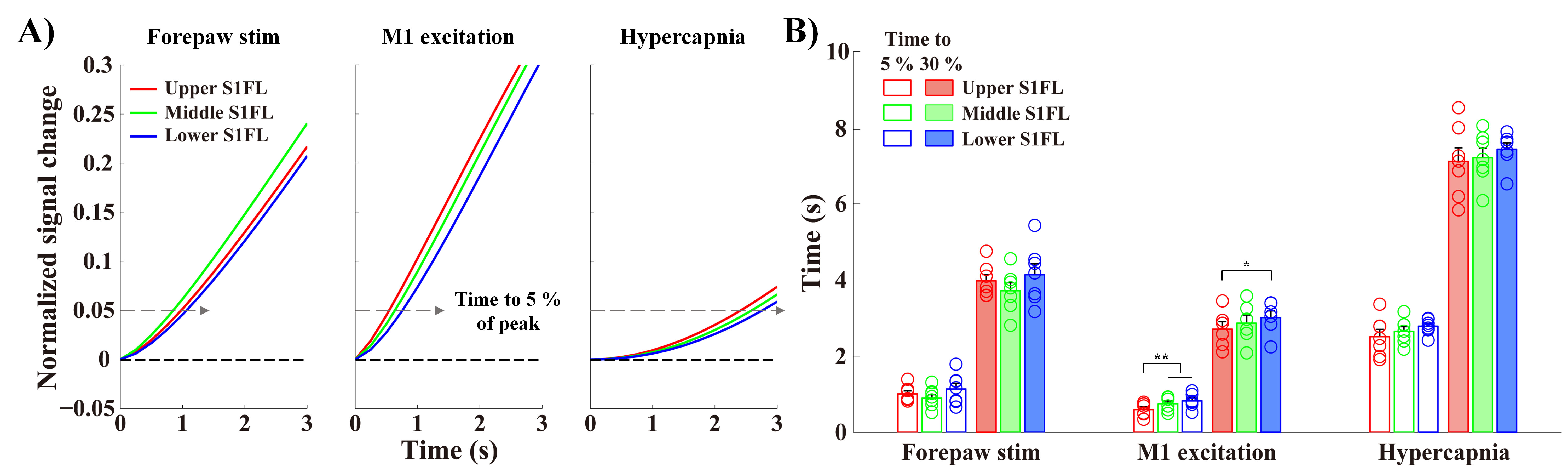
Figure 3. Layers receiving synaptic inputs show the earliest BOLD responses.
A-B) The fitting curves were normalized to the peak (showing from 0 s to 3 s after stimulus onset), and the times to reach 5 % and 30 % of the peak were then measured in the S1FL layer-specific ROIs. The order of early BOLD responses was Middle → Upper → Lower for forepaw stimulation and Upper → Middle → Lower for M1 excitation and hypercapnia.
error bars in B, SEM; colored circles in B, individual animal data; * and ** in B, p < 0.05 and p < 0.01, respectively (repeated ANOVA followed by Tukey post hoc test).