Vahid Khalilzad Sharghi1, Eric Maltbie1, Wen-Ju Pan1, Shella Keilholz1, and Kaundinya Gopinath2
1Biomedical Engineering, Emory University/Georgia Institute of Technology, Atlanta, GA, United States, 2Department of Radiology & Imaging Sciences, Emory University, Atlanta, GA, United States
1Biomedical Engineering, Emory University/Georgia Institute of Technology, Atlanta, GA, United States, 2Department of Radiology & Imaging Sciences, Emory University, Atlanta, GA, United States
Suppression of cortical slow rhythms led to strong reductions in the
amplitudes of quasi-periodic patterns (QPPs). On the other hand, functional
connectivity in canonical brain function networks increased significantly.
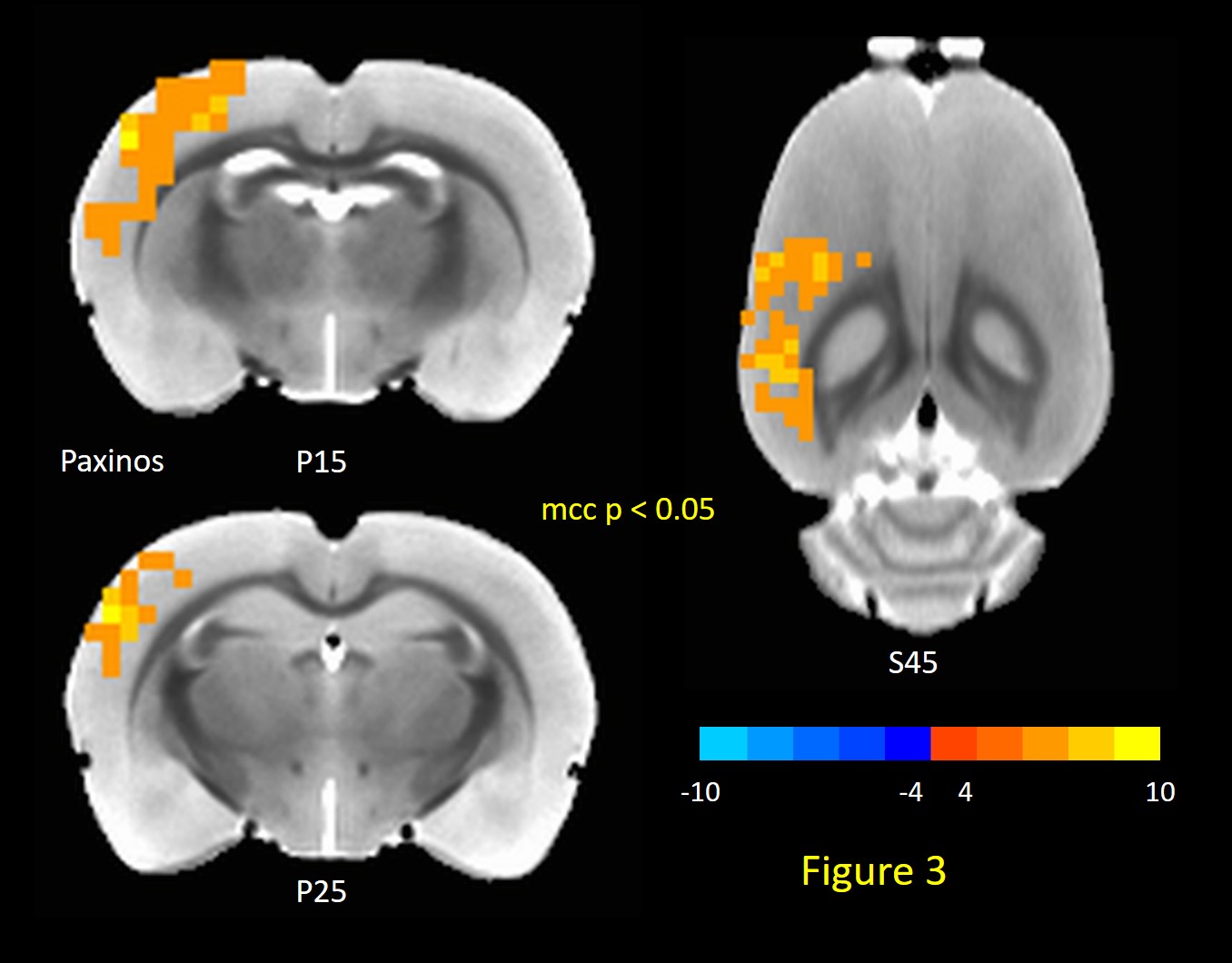
Fig.3: TTA-P2 vs Baseline t-statistic maps
highlighting regions with enhanced FC to the ROI encompassing all right
hemisphere auditory cortex regions after TTA-P2 administration. The
slice-location co-ordinates are in Paxinos space 16,17. Left-hemisphere
is on the left-hand side of the maps.
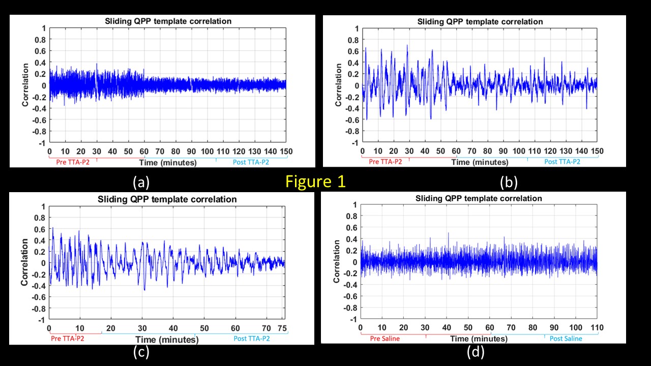
Fig.1: The evolution of the strengths of QPPs with time
assessed with spatio-temporal correlation of the fMRI time-series with
corresponding QPP template. Examples from (a-c) three rats after systematic
administration of TTA-P2; and one rat (d) after systematic administration of
Vehicle.