Seung Eun Lee1, Joon-Yong Jung1, and Dongyeob Han2
1Seoul St. Mary’s Hospital, Seoul, Korea, Republic of, 2Siemens Healthineers, Seoul, Korea, Republic of
1Seoul St. Mary’s Hospital, Seoul, Korea, Republic of, 2Siemens Healthineers, Seoul, Korea, Republic of
In ex vivo experiment
and clinical study, we demonstrated that SFF map generated by multicomponent
MRF framework can quantify synovial fluid in small cartilage defect, and
provides direct information on cartilaginous water content.
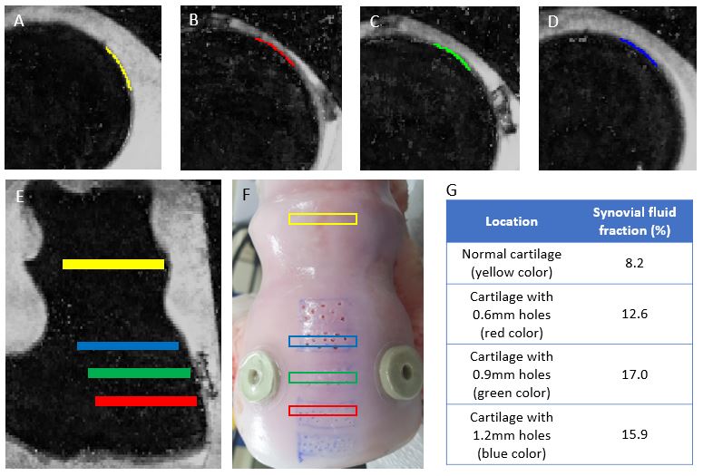
Figure 1. Cartilage
segmentation on bovine cartilage in normal cartilage (A), cartilage with 0.6mm
holes (B), 0.9mm holes (C) and 1.2mm holes (D). (E, F) Segmentation area
indicated with colors in sagittal plane and leg specimen. (G) Result of
cartilaginous fluid fraction in each location.
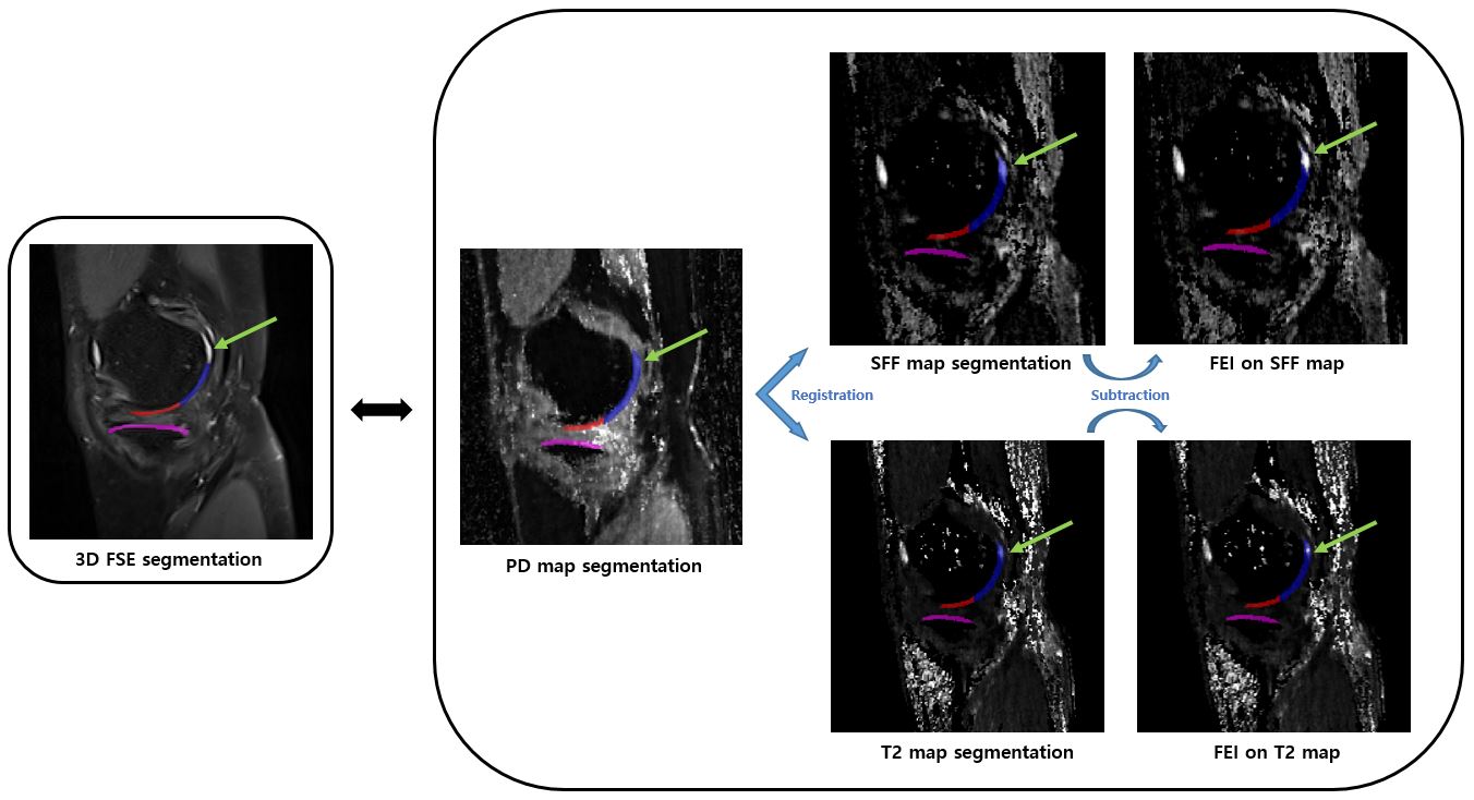
Figure 3. Knee
cartilage segmentations of 3D FSE image and PD map in osteoarthritis patient. A
cartilage defect in posterior medial femoral condyle is demonstrated in 3D FSE
image (green arrow) and shows good correlation with fluid exclusion image (FEI)
on SFF map, better than FEI on T2 map.