Iman Khodarahmi1, Inge Manuela Brinkmann2, Dana Lin1, Mary Bruno1, Patricia Johnson1, Florian Knoll1, Mahesh Bharath Keerthivasan2, Hersh Chandarana1, and Jan Fritz1
1Department of Radiology, New York University School of Medicine, New York, NY, United States, 2Siemens Medical Solutions USA Inc., Malvern, PA, United States
1Department of Radiology, New York University School of Medicine, New York, NY, United States, 2Siemens Medical Solutions USA Inc., Malvern, PA, United States
Metal artifacts from hip arthroplasty implants are
substantially smaller at 0.55T than clinical 1.5T, holding great promise for improved
metal artifact reduction MRI of hip arthroplasty implants. Our preliminary
results suggest clinically viable sequence acquisition times of ≤ 6-min.
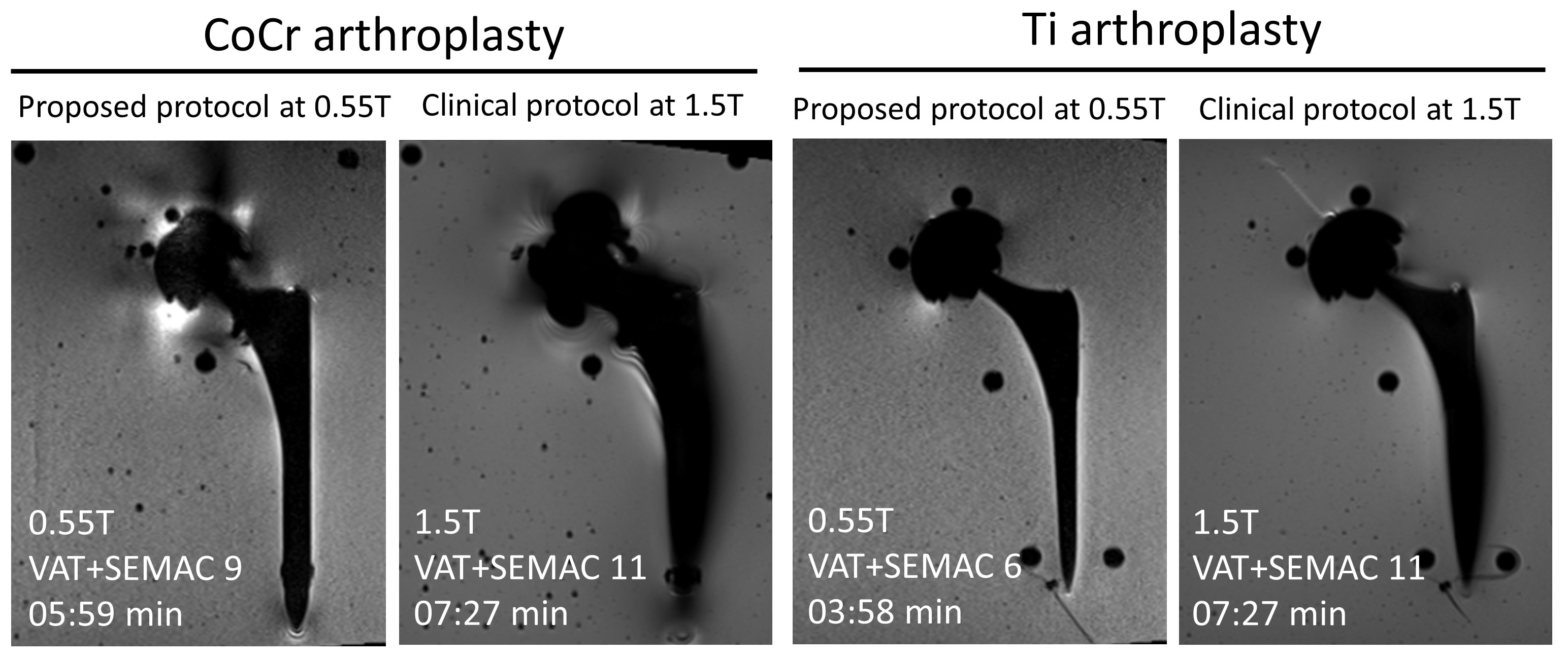
Figure 3: Coronal MR images of cobalt-chromium and titanium-on-ceramic
hip arthroplasty implant systems using the clinical protocol at 1.5T and the proposed
protocol at 0.55T. Despite higher SEMAC encoding steps at 1.5T, metal artifacts
are substantially smaller at 0.55T.
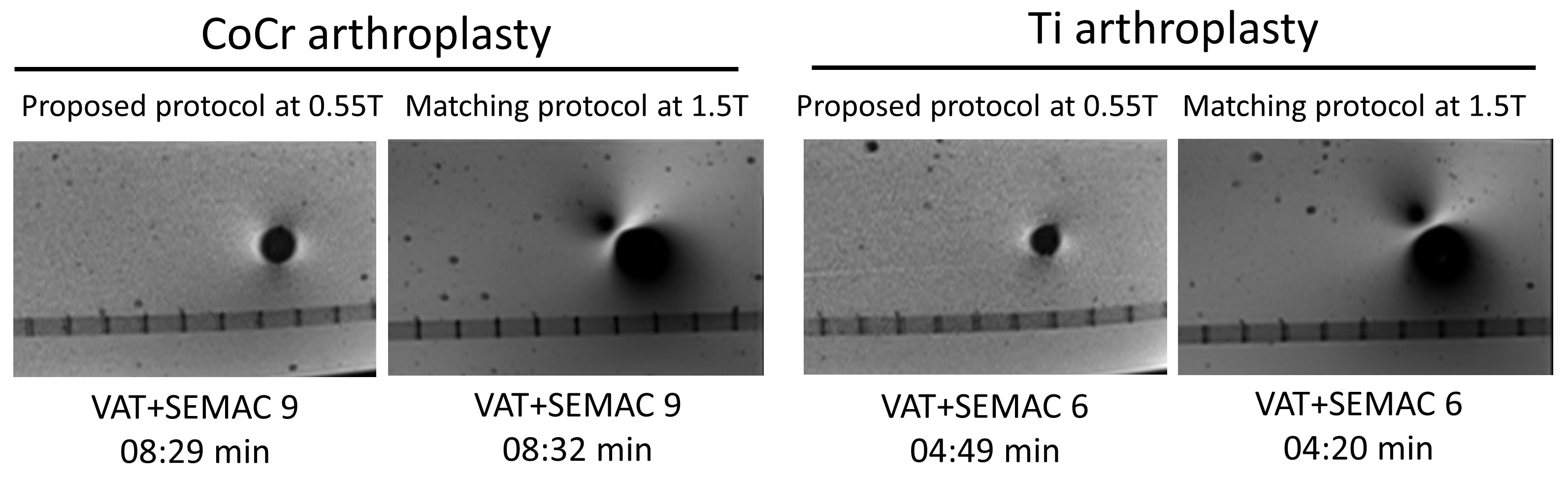
Figure 4: Axial MR images of cobalt-chromium and
titanium-on-ceramic hip arthroplasty implant systems using matched protocols at
1.5T and 0.55T. Metal artifacts are substantially smaller at 0.55T.