Cihat Eldeniz1, Udayabhanu Jammalamadaka1, Gary B. Skolnick2, Paul K. Commean1, Kamlesh B. Patel2, and Hongyu An1
1Mallinckrodt Institute of Radiology, Washington University in St. Louis, St. Louis, MO, United States, 2Division of Plastic and Reconstructive Surgery, Washington University in St. Louis, St. Louis, MO, United States
1Mallinckrodt Institute of Radiology, Washington University in St. Louis, St. Louis, MO, United States, 2Division of Plastic and Reconstructive Surgery, Washington University in St. Louis, St. Louis, MO, United States
CT may cause
cancer due to ionizing radiation. MRI is vulnerable to motion. Sedation reduces
motion, but can be harmful. In order to replace CT and skip sedation, we
developed an MRI scheme that is robust to motion and also performs motion
correction.
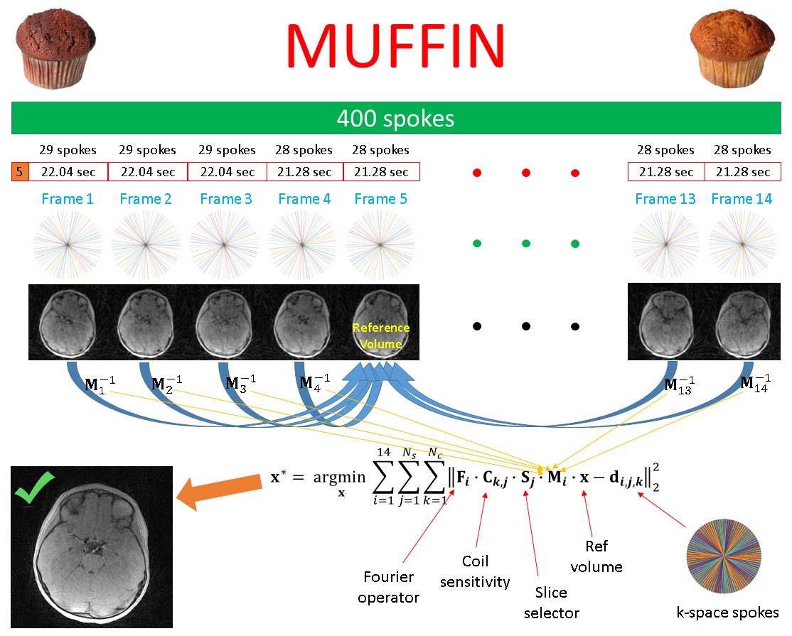
MUFFIN
processing. The first 5 spokes were discarded to reach a steady state. The rest
was split into 14 frames. 40% apodization was a good tradeoff between
resolution and undersampling-related artifacts. The density compensation
function was V-shaped. After determining the reference frame (by picking the
closest rotation vector to the mean rotation vector), all transformations to
and from the reference frame were used in the optimization to obtain the
corrected volume.
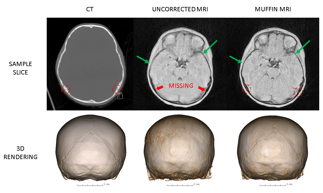
Top row: A sample slice for Patient 1 acquired with CT and MRI. The
dashed red circles indicate the sutures (quite fine structures, and hence
difficult to see without looking closely).The uncorrected image is missing both
sutures. The green arrows exemplify the detail and sharpness recovered by
MUFFIN. Bottom row: 3D renderings. The uncorrected one is barely showing any
sutures, while the similarity between the CT skull and the MUFFIN MRI skull is
striking.