Kevin Matthew Koch1, Mohammad Zarenia2, V. Emre Arpinar2, L Tugan Muftuler3, Alyssa Joy Schnorenberg 4, Joshua Leonardis4, Brooke Slavens4, and Andrew S Nencka2
1Medical College of Wisconsin, Milwaukee, WI, United States, 2Radiology, Medical College of Wisconsin, Milwaukee, WI, United States, 3Neurosurgery, Medical College of Wisconsin, Milwaukee, WI, United States, 4University of Wisconsin, Milwaukee, Milwaukee, WI, United States
1Medical College of Wisconsin, Milwaukee, WI, United States, 2Radiology, Medical College of Wisconsin, Milwaukee, WI, United States, 3Neurosurgery, Medical College of Wisconsin, Milwaukee, WI, United States, 4University of Wisconsin, Milwaukee, Milwaukee, WI, United States
4D dynamic MRI is combined with a slab-to-volume boundary-based registration to high resolution static images in navigating carpal bones through unconstrained motion. Sample temporal kinematic carpal profiles constructed with this method are analyzed for viability in future analyses.
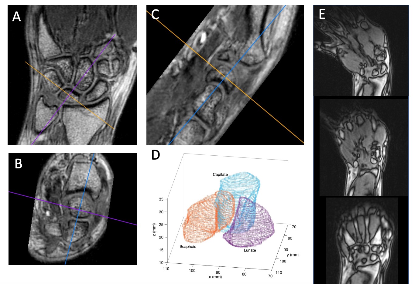
Figure 1: A-C) Orthogonal imaging planes through the carpal bones of a 3D SPGR acquisition utilized to segment the bones of interest. Segmentation was performed manually and takes into account the cortical bone gap boundary on the images. D) Resulting point cloud of boundary points derived from the high-resolution segmentations. E) Sample slices of 3D dynamic SPGR images utilized to track the static segmentations and the points of interest identified on those segmentations. Kinematic profiles are derived from the tracked points on the static segmentations.
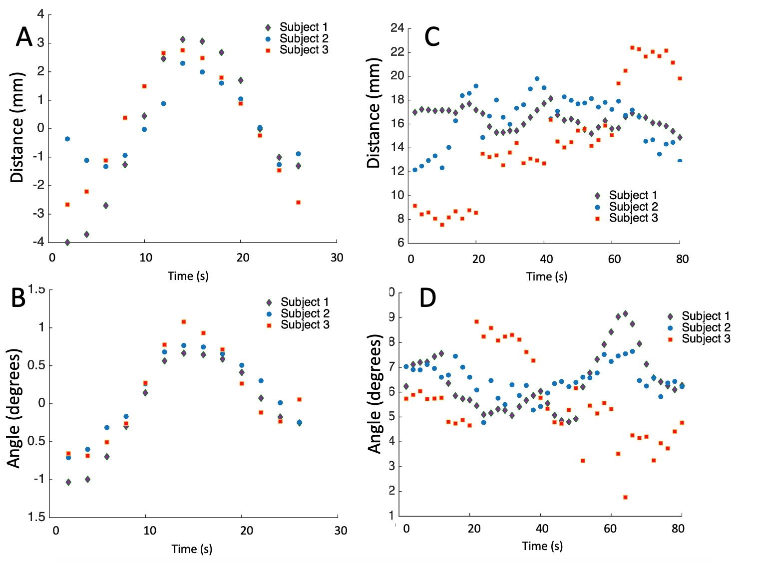
Figure 4: Exemplary kinematic profiles from the study cohort. A) Scaphoid-Capitate center of mass distance and B) Scaphoid-Capitate center of mass angle. For reference and to demonstrate the impact of the basic processing steps applied to generate these profiles, the raw input data for these profiles is displayed in C) scaphoid-capitate center of mass distance and D) scaphoid capitate center of mass angle.