Preety Krishnan1, Tejas J Shah2, Akshay Godkhindi2, Rupsa Bhattacharjee3, Stanley Kovil Pichai3, Ajay Krishnan1, Bharat Dave1, and Indrajit Saha3
1Stavya Spine Research Institute, Ahmedabad, India, 2MR, Philips Innovation Campus, Bangalore, India, 3Philips India Limited, Gurgaon, India
1Stavya Spine Research Institute, Ahmedabad, India, 2MR, Philips Innovation Campus, Bangalore, India, 3Philips India Limited, Gurgaon, India
The
proposed logistic regression model based on 9 texture features, highlighted in Figure
3 in green, can be used clinically on 1.5T
systems on T1W images to detect osteoporosis in Spine.
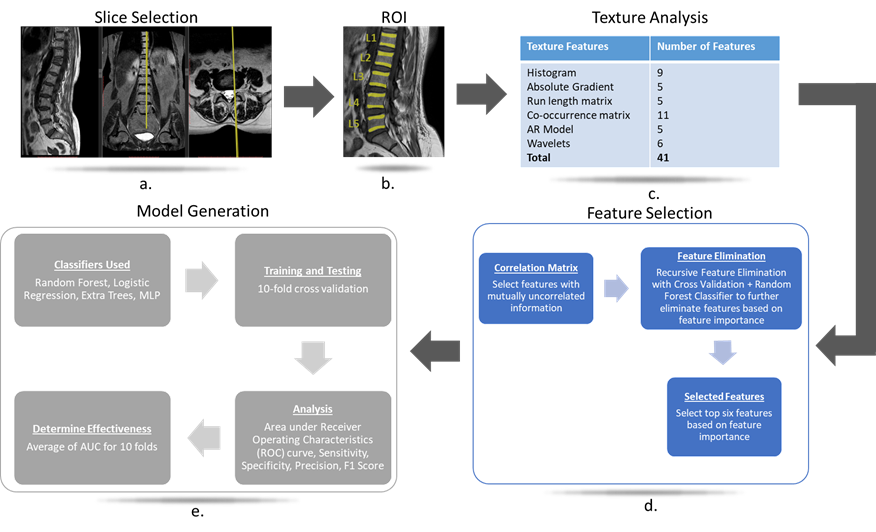
Figure 1: Process
for automated texture analysis based classification. Location of the sagittal
slice selected for subsequent analysis is shown in a., The regions of interest
(ROIs) selected manually for L1-L5 vertebrae are shown in b., the classes of
texture features computed are shown in c., the process of feature selection is
shown in d. and the steps for generation and selection of classifier models for
classification of cases into osteoporotic or non-osteoporotic is shown in e.
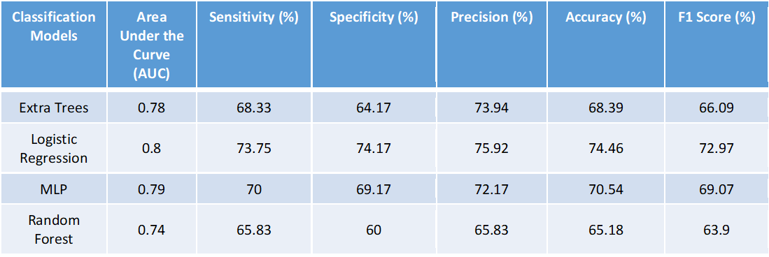
Table 2: ROC analysis parameters for
different classifiers indicating their respective effectiveness