Brandon Clinton Jones1,2, Hyunyeol Lee1, Shaowei Jia1,3, Anna Feng1, Snehal S Shetye4, Hee Kwon Song1, Felix Werner Wehrli1, and Chamith Sudesh Rajapakse1,4
1Radiology, University of Pennsylvania, Philadelphia, PA, United States, 2Bioengineering, University of Pennsylvania, Philadelphia, PA, United States, 3Biomedical Science and Medical Engineering, Beihang University, Beijing, China, 4Orthopaedic Surgery, University of Pennsylvania, Philadelphia, PA, United States
1Radiology, University of Pennsylvania, Philadelphia, PA, United States, 2Bioengineering, University of Pennsylvania, Philadelphia, PA, United States, 3Biomedical Science and Medical Engineering, Beihang University, Beijing, China, 4Orthopaedic Surgery, University of Pennsylvania, Philadelphia, PA, United States
Pore water
content, total water content, and porosity index were all associated with
whole-bone stiffness and mineral density in cadaveric proximal femora. Cortical
bone water measures may therefore provide useful information on cortical bone
health.
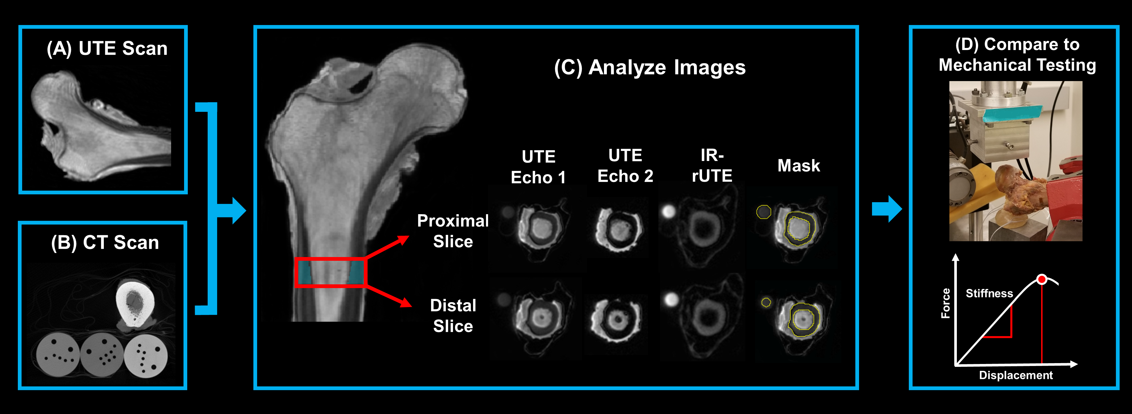
Flow chart illustrating workflow for the study. (A) representative
sagittal UTE image. (B) axial slice of CT scan with the 3 calcium calibration rods. (C) 1-cm cortical analysis mask region which is chosen just inferior
to the lesser trochanter. The proximal and distal slices of the UTE sequences are shown. Note higher signal within the cortical bone in
the first than in the second echo image. Soft tissue is suppressed in the IR
sequence, retaining only bound water signal from the cortical bone and the
calibration sample. (D) overview of the mechanical testing
methodology.
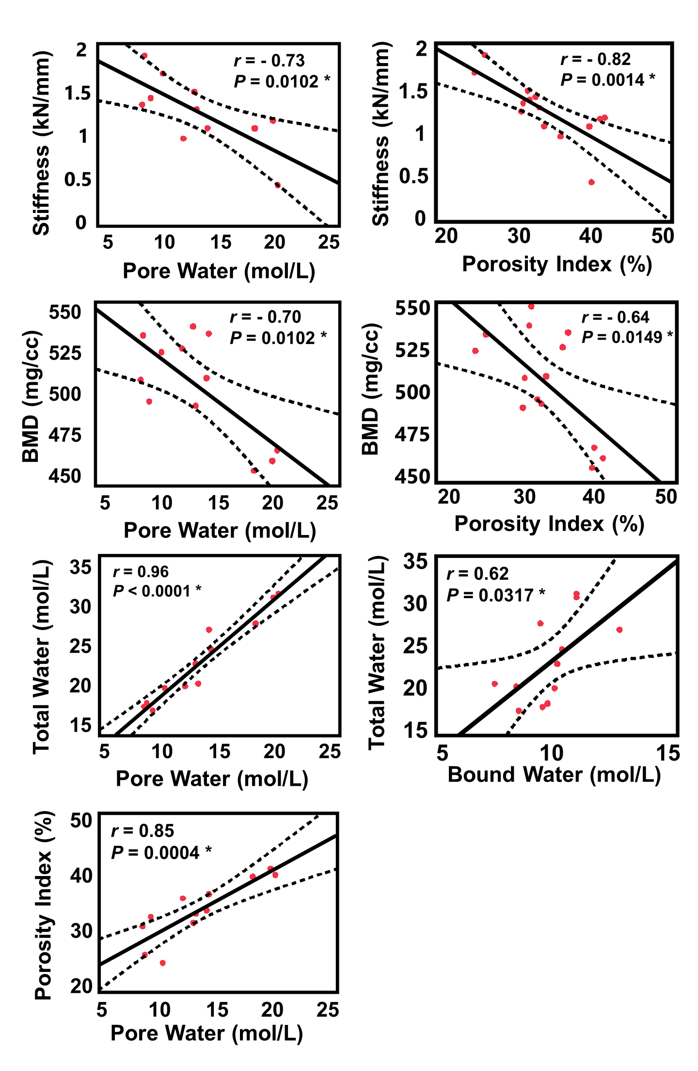
Correlation plots between imaging parameters and whole-bone stiffness.
Error clouds indicate 95% confidence intervals. Asterisks indicate significant correlations.
Pore water and porosity index, which are surrogates of cortical porosity, were negatively
correlated to stiffness and mineral density. Pore water content and porosity index
were also strongly positively correlated with each other. Pore water concentrations
were greater than bound water concentrations and thus contributed more to the
total water content.