Sophia Kronthaler1, Christof Boehm1, Peter Börnert2, Ulrich Katscher2, Kilian Weiss3, Marcus R. Makowski1, Benedikt J. Schwaiger1, Alexandra S. Gersing1, and Dimitrios C. Karampinos1
1Department of Diagnostic and Interventional Radiology, School of Medicine, Technical University of Munich, Munich, Germany, 2Philips Research Laboratory, Hamburg, Germany, 3Philips Healthcare, Hamburg, Germany
1Department of Diagnostic and Interventional Radiology, School of Medicine, Technical University of Munich, Munich, Germany, 2Philips Research Laboratory, Hamburg, Germany, 3Philips Healthcare, Hamburg, Germany
The proposed methodology enables single-point Dixon and SW imaging the simultaneous assessment of vertebral fractures and edema of the thoracolumbar spine from a single-TE UTE sequence.
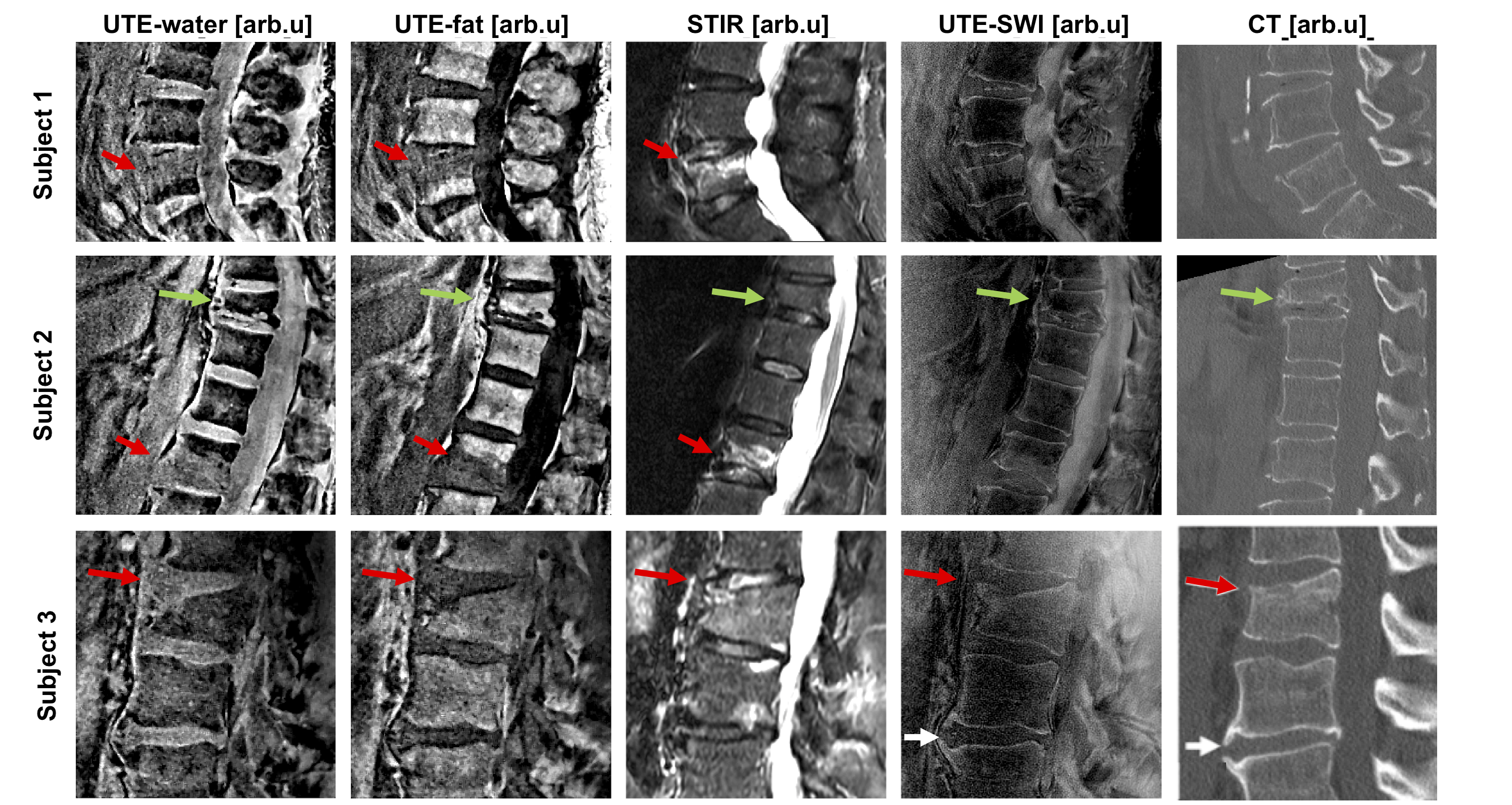
Figure 5: Comparison of spDixon, STIR, UTE-SWI and CT in three patients with acute vertebral fractures (red arrows) and bone marrow edema (S1 and S2). The edema and fracture line were visible in the spDixon Water–fat images and the SWI images. The second subject showed signs of a chronic vertebral fracture (green arrow), without signal alterations neither in the STIR nor the UTE image. The third subject showed signs of an acute vertebral fracture such as a compaction zone (red arrows) as well as ventral and a small dorsal spondylophytes (white arrows) one level lower.
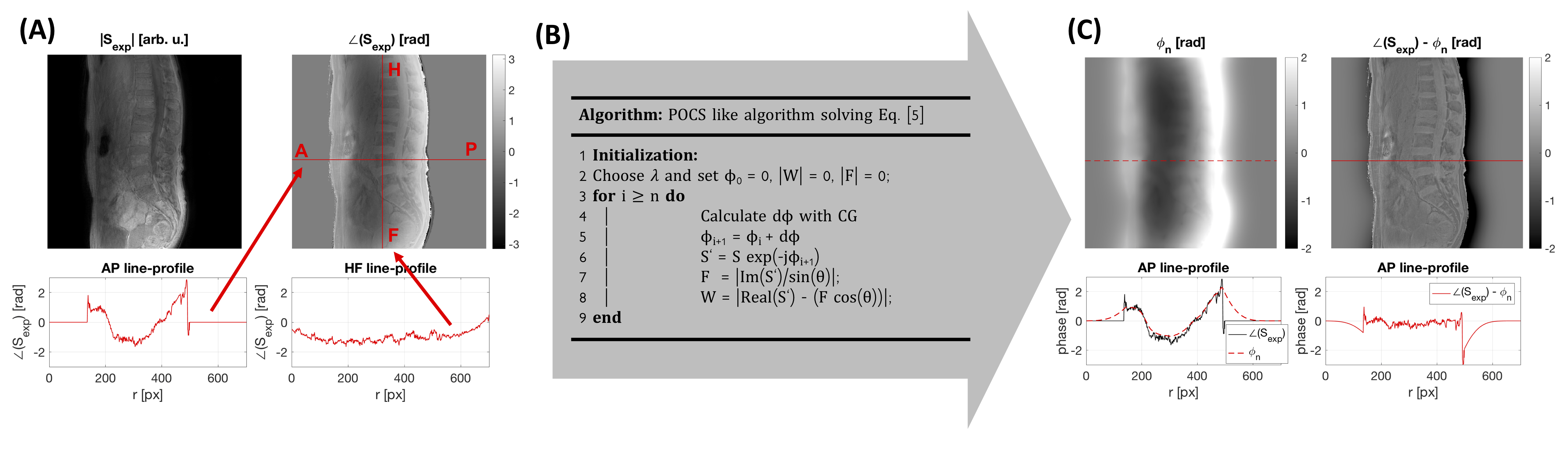
Figure 1: spDixon processing pipeline: A) The UTE phase $$$\angle\left(S_{\text{exp}}\right)$$$ contained contributions from the B1 phase which varied along AP and RL (x-y-plane). B) A POCS-like algorithm was used to solve Equation [4]. The update dϕ was calculated using conjugate gradients (CG). C) After n-update steps the phase error ϕn was estimated. In the corrected UTE phase ϕ0 – ϕn unwanted phase components and the B1 phase contribution were removed.