Yasuhisa Kurata1, Mizuho Nishio1, Yusaku Moribata2, Aki Kido1, Yuki Himoto1, Koji Fujimoto3, Masahiro Yakami2, Sachiko Minamiguchi4, Masaki Mandai5, and Yuji Nakamoto1
1Diagnostic Imaging and Nuclear Medicine, Kyoto university hospital, Kyoto, Japan, 2Preemptive Medicine and Lifestyle-Related Disease Research Center, Kyoto university hospital, Kyoto, Japan, 3Real World Data Research and Development, Graduate School of Medicine Kyoto University, Kyoto, Japan, 4Diagnostic Pathology, Kyoto university hospital, Kyoto, Japan, 5Gynecology and Obstetrics, Kyoto university hospital, Kyoto, Japan
1Diagnostic Imaging and Nuclear Medicine, Kyoto university hospital, Kyoto, Japan, 2Preemptive Medicine and Lifestyle-Related Disease Research Center, Kyoto university hospital, Kyoto, Japan, 3Real World Data Research and Development, Graduate School of Medicine Kyoto University, Kyoto, Japan, 4Diagnostic Pathology, Kyoto university hospital, Kyoto, Japan, 5Gynecology and Obstetrics, Kyoto university hospital, Kyoto, Japan
The model developed in this study has achieved high-accuracy automatic segmentation of endometrial cancer on MRI using a convolutional neural network for the first time. Using multi-sequence MR images were important for high accuracy segmentation.
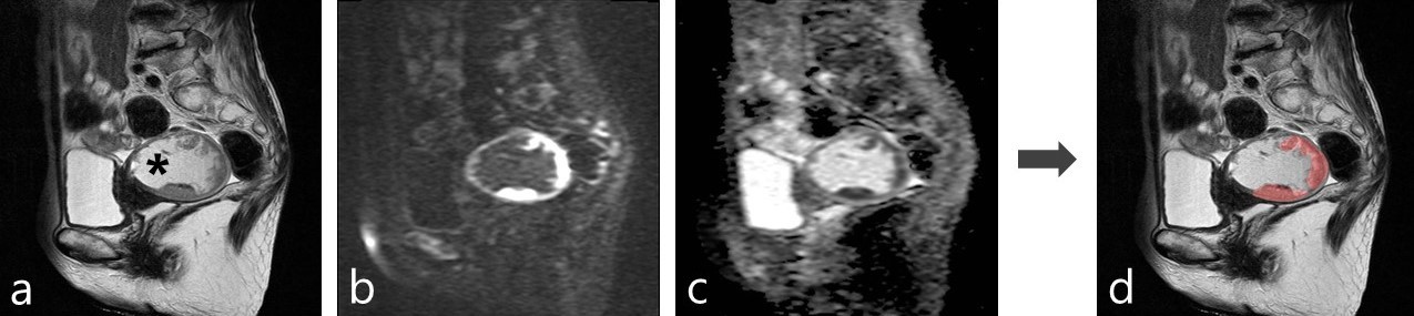
Figure 5: A representative case of the automatic segmentation
a: T2-weighted image
b: diffusion-weighted image (b=1000 s/mm2)
c: apparent diffusion coefficient map
d: A result of automatic segmentation of endometrial cancer overlaid on T2-weighted image
The tumor was well segmented despite the presence of hematometra (a:*) (Dice similarity coefficient=0.808).
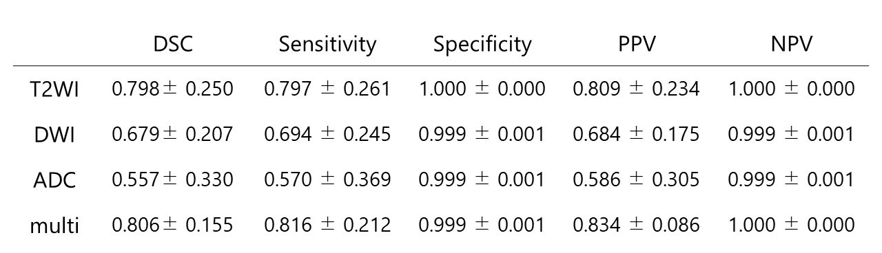
Figure 3: The segmentation accuracy of our model for the test datasets with each MRI sequences as input data
Data are presented with mean±standard deviation
T2WI: T2-weighted image
DWI: diffusion-weighted image
ADC: apparent diffusion coefficient
Multi: T2WI, DWI, and ADC map
DSC: Dice similarity coefficient
PPV: positive predictive value
NPV: negative predictive value
