Thomas Welton1,2, Septian Hartono3, Yao-Chia Shih3, Samuel Y-E Ng1, Nicole S Y Chia1, Weiling Lee3, Say Lee Chong3, Eng-King Tan1,2,3, Ling-Ling Chan1,2,3, and Louis CS Tan1,2
1National Neuroscience Institute, Singapore, Singapore, 2Duke-NUS Medical School, Singapore, Singapore, 3Singapore General Hospital, Singapore, Singapore
1National Neuroscience Institute, Singapore, Singapore, 2Duke-NUS Medical School, Singapore, Singapore, 3Singapore General Hospital, Singapore, Singapore
We found
elevated mean kurtosis in specific basal ganglia regions, which was maintained
over two years and negatively correlated with worsening motor function in
Parkinson’s disease.
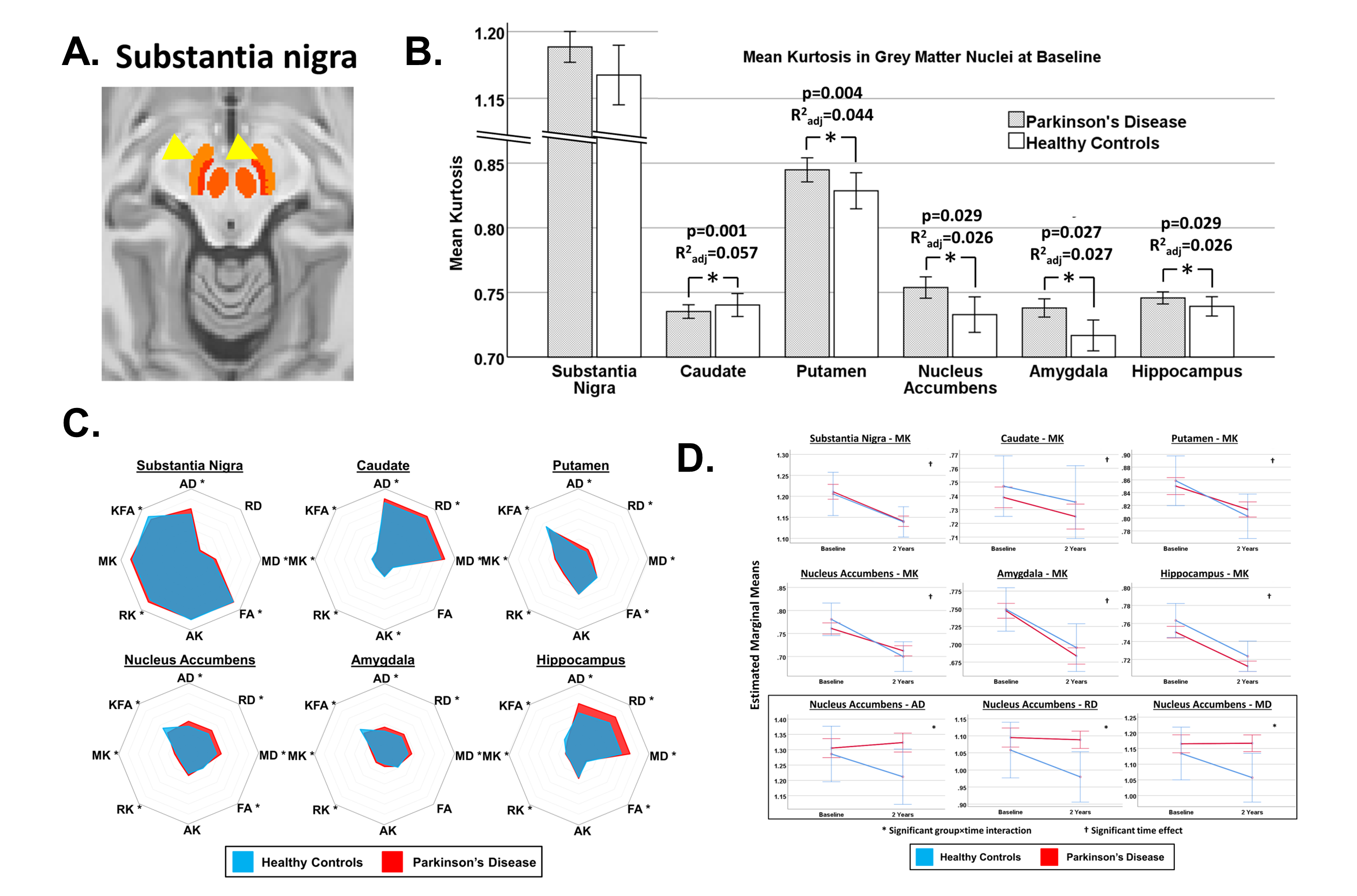
Figure
1. (A) Substantia nigra mask (orange region) from the CIT168 atlas. (B) Mean
kurtosis in each region of interest compared between groups. Bars show the raw
uncorrected data while p-values and adjusted R2 values given are for
the corrected model controlling for age, sex and education. (C) Radial plots of
diffusion kurtosis indices for each region and both groups. (D) Plots of the estimated marginal means from
repeated-measures ANOVA. Error bars show 95%CI. * Significant for p<0.05.