Matthew Kim1, Denise Zhong1, Moss Y Zhao2, David Y.T Chen3, David D Shin4, Greg Zaharchuk2, and Audrey P. Fan1,5
1Department of Biomedical Engineering, University of California, Davis, Davis, CA, United States, 2Department of Radiology, Stanford University, Stanford, CA, United States, 3Department of Medical Imaging, Taipei Medical University-Shuan-Ho Hospital, New Taipei City, Taiwan, 4General Electric Healthcare, San Ramon, CA, United States, 5Department of Neurology, University of California Davis, Davis, CA, United States
1Department of Biomedical Engineering, University of California, Davis, Davis, CA, United States, 2Department of Radiology, Stanford University, Stanford, CA, United States, 3Department of Medical Imaging, Taipei Medical University-Shuan-Ho Hospital, New Taipei City, Taiwan, 4General Electric Healthcare, San Ramon, CA, United States, 5Department of Neurology, University of California Davis, Davis, CA, United States
(1) Multi-parametric R2’ values are reproducible in repeat sessions 1-2 weeks apart within cortical vascular territories; (2) Whole brain R2’ maps present higher values in Moyamoya patients compared to healthy volunteers, indicating abnormally high oxygen extraction fraction (OEF).
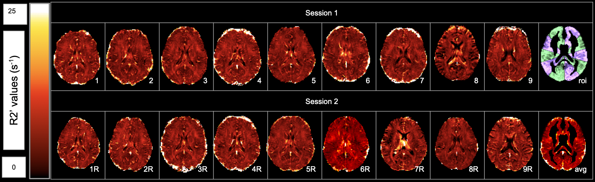
Figure 1 - R2’ maps of each healthy volunteer. 1-9 are from initial scans of each volunteer, 1R-9R are from repeat scans taken 1-2 weeks after the first scan. Avg represents combined average R2’ map of all maps in Session 1 and Session 2. ROI represents the regions of interest used to analyze R2’ values.
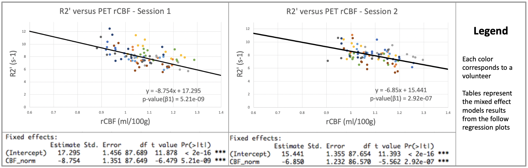
Figure 3 - R2’ and rCBF at baseline for Healthy control between initial scans for each volunteer and their mixed effects model output below (Session 1) and repeat scans for each volunteer taken a week or more from the initial scans and their mixed effects model output below (Session 2).