Lingceng Ma1,2, Qingjia Bao1, Ricardo P. Martinho1, Zhong Chen2, and Lucio Frydman1
1Department of Chemical and Biological Physics, Weizmann Institute of Science, Rehovot, Israel, 2Department of Electronic Science, Xiamen University, Xiamen, China
1Department of Chemical and Biological Physics, Weizmann Institute of Science, Rehovot, Israel, 2Department of Electronic Science, Xiamen University, Xiamen, China
Fast T1 mapping methods based on
subspace-constrained reconstructions of jointly sparse-sampled domains, are
proposed and shown to efficiently deliver maps with either multiple T1
contrasts or T1 values, on both human or animal MRI scans, with remarkable
accelerations.
Figure
5.
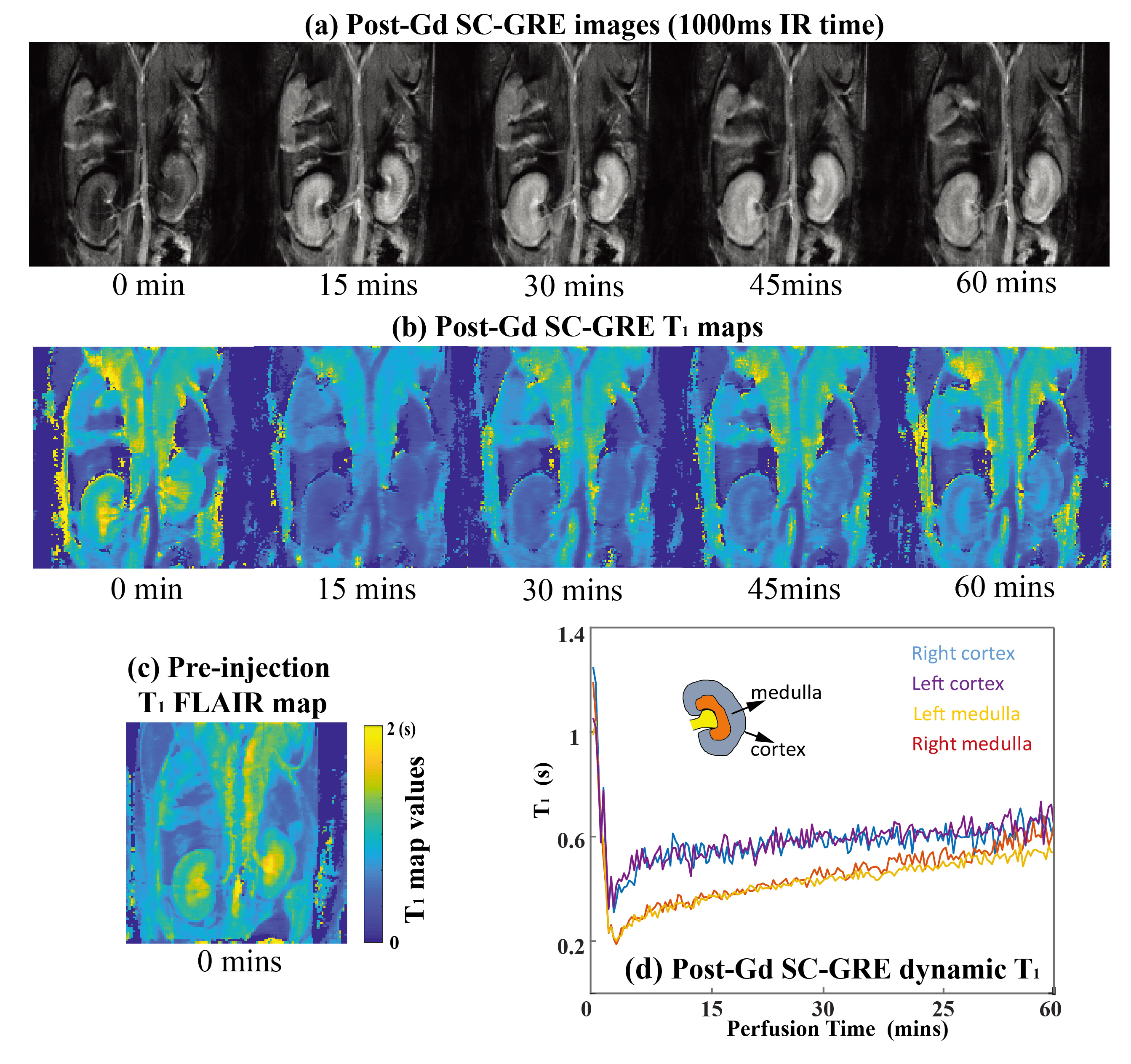
Real-time T1 renal mapping of a live mouse upon injection of a Gd-DTPA bolus
(0.1mmol/kg, mice weight = 25g). (a) SC IR GRE T1 images reflecting
an IR=1s, collected over the course of one hour. (b) Dynamic SC IR GRE T1 maps collected over one hour. (c) T1 FLAIR maps
acquired before the dynamic SC IR GRE T1 mapping. (d) Dynamic T1 values extracted from both
kidneys' cortex and medulla regions. The parameters of T1-FLAIR T1 mapping and SC-GRE T1 mapping are the same as the ones used in Fig.4.
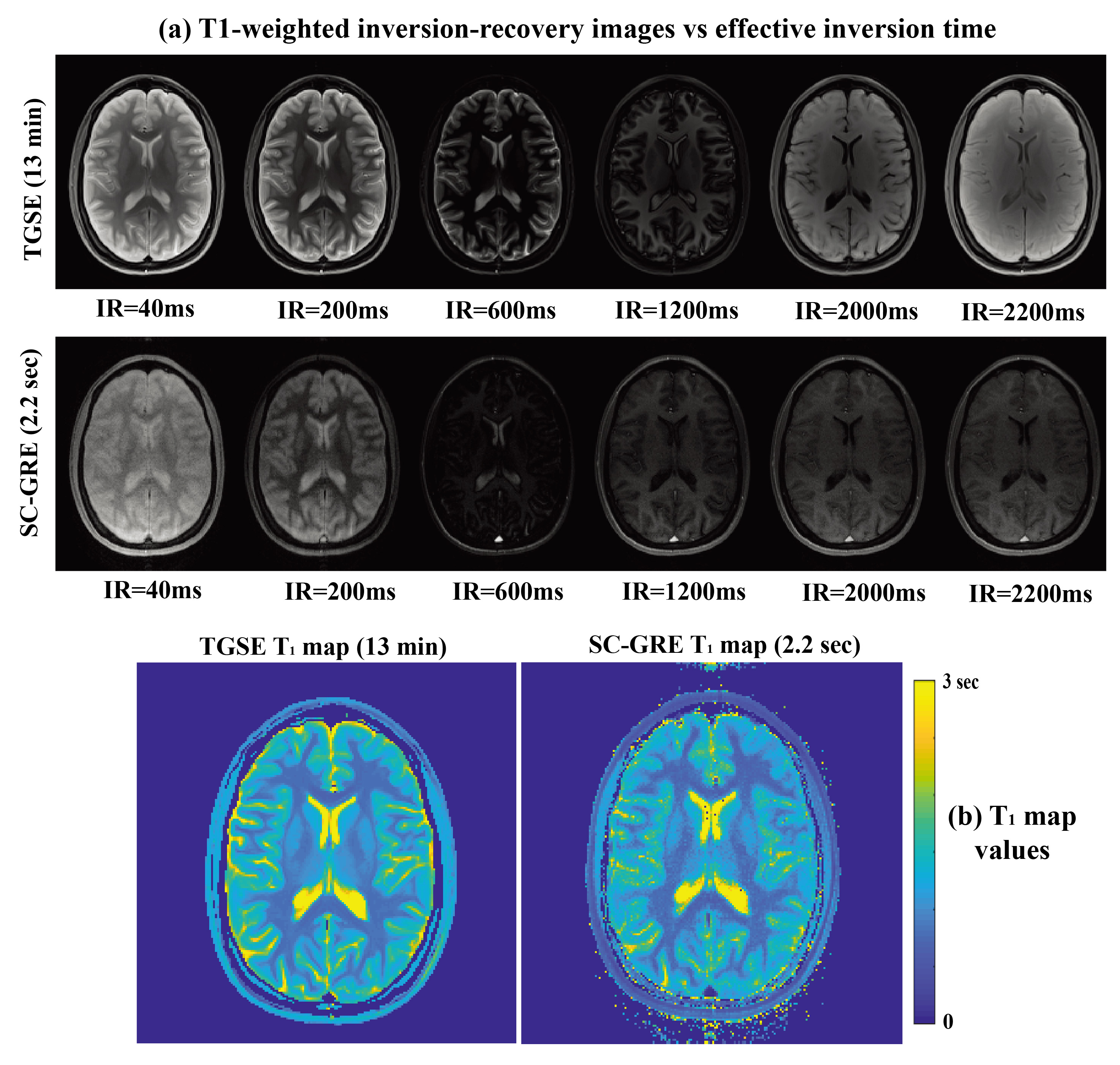
Figure 2. SC IR GRE and TGSE T1 weighting and mapping experiments on a human
brain. (a) Inversion-recovery weighted TGSE and SC GRE images. (b) T1 maps afforded for both method. TGSE T1 mapping
parameters: 1.15×1.15×3mm3, TR = 6s, 2 averages, IR times =[0.04,
0.20, 0.60,1.20, 2.00, 3.00]s, 7 spin echo trains with 3 GRE echoes, total
acquisition time=13mins. SC IR GRE parameters: 1.15×1.15×3mm3,
Flip Angle excitation pulse (FA) = 10o, TR = 10ms, 219 GRE readouts after the inversion pulse, total acquisition time = 2.2s. Reconstruction was as in Fig. 1d, with a K = 2 SVD
subspace base.