Rosa Sanchez Panchuelo1, Olivier Mougin1, Robert Turner1,2, and Susan Francis1,3
1Sir Peter Mansfield Imaging Centre, UP, University of Nottingham, Nottingham, United Kingdom, 2Max Planck Institute for Human Cognitive and Brain Sciences, Leibzig, Germany, 3NIHR Nottingham Biomedical Research, University of Nottingham, Nottingham, United Kingdom
1Sir Peter Mansfield Imaging Centre, UP, University of Nottingham, Nottingham, United Kingdom, 2Max Planck Institute for Human Cognitive and Brain Sciences, Leibzig, Germany, 3NIHR Nottingham Biomedical Research, University of Nottingham, Nottingham, United Kingdom
We present a model that effectively removes magnetization transfer contributions of spectrally-selective fat-suppression pulses from measured T1, hence providing accurate T1 quantification in multi-slice IR-EPI with respect to single slice IR-EPI measures.
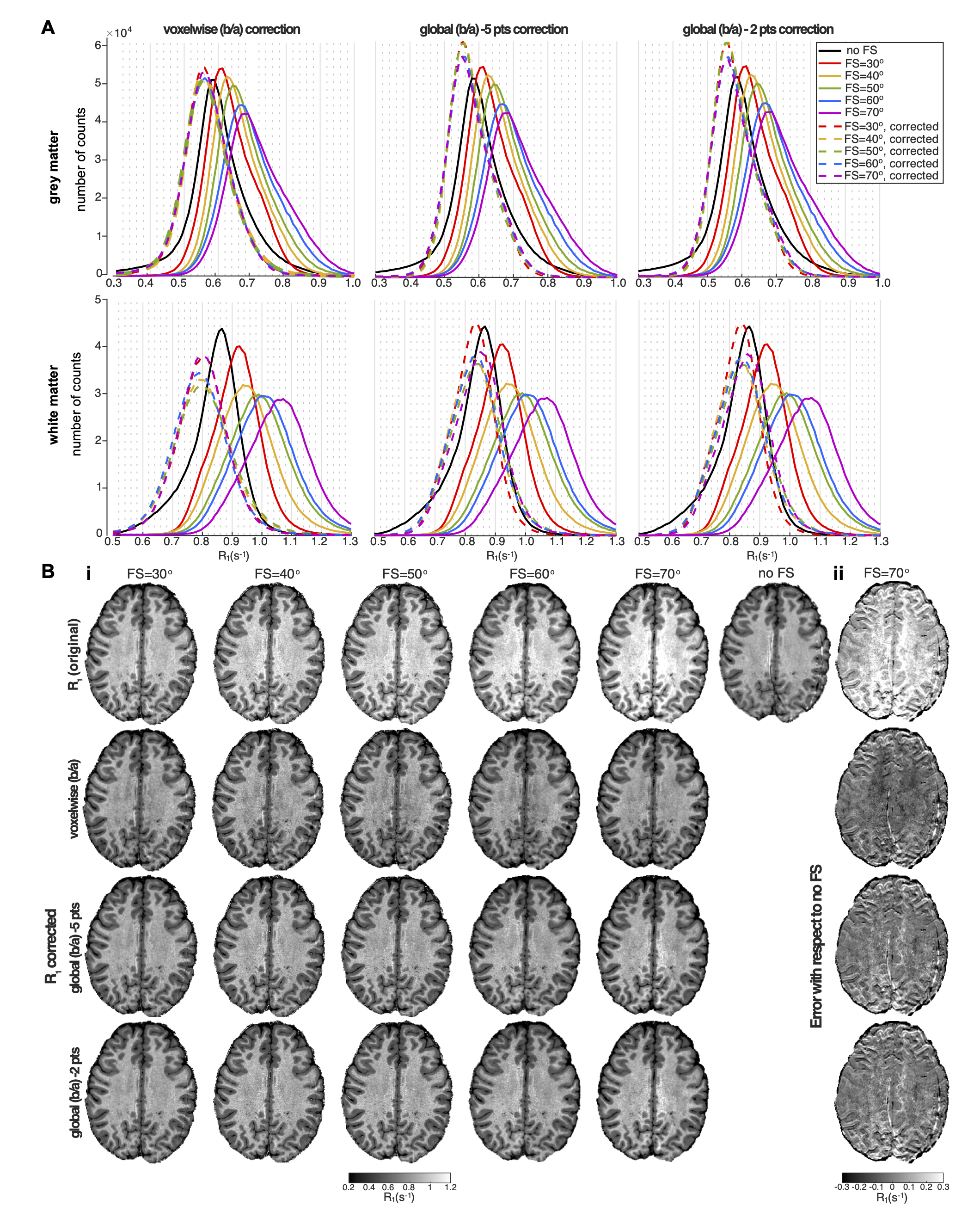
Figure 3: (A) R1-histograms for different FS levels
(black: no FS) before (solid) and after (dash) correction using different
correction methods; voxel-wise (b/a)-correction (left) and global-(b/a) correction using full (5pts)
data set (middle) and reduced (2 pts) data set (right). (B) (i) Original and corrected R1 maps. (ii) Variance of corrected R1 map (SPIR FA=70o) wrt no FS.
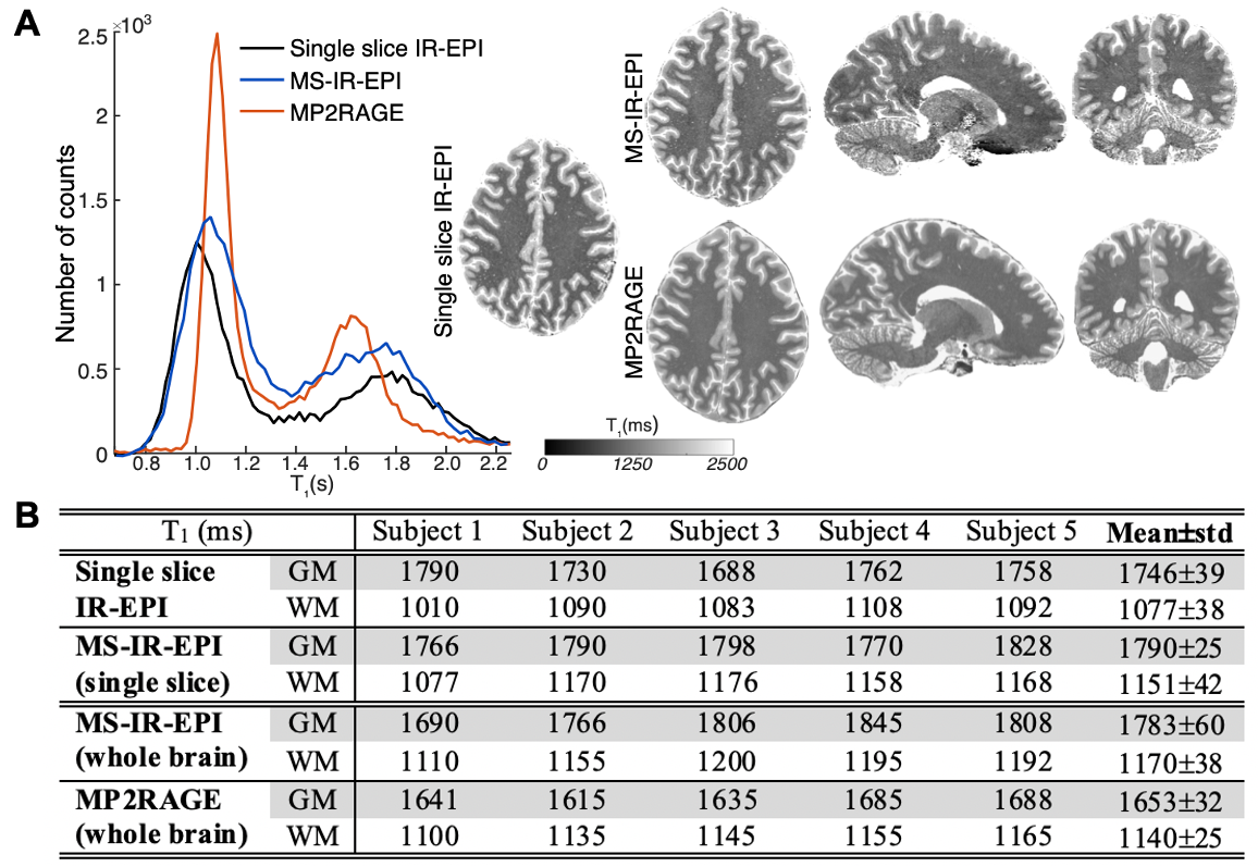
Figure 5: (A) T1-histograms for a single slice (shown) for standard single slice IR-EPI
(black line), MP2RAGE (orange) and MS-IR-EPI after correction (blue). Sagittal and coronal views from whole brain
dataset also shown for MS-IR-EPI (top raw) and MP2RAGE (bottom). (B) T1-values of histogram peak for data
acquired with standard single slice IR-EPI, MS-IR-EPI with fat suppression
(both same slice as the standard IR-EPI and whole brain) and whole brain
MP2RAGE.