Jianshu Chi1, Victor Han1, and Chunlei Liu1,2
1Electrical Engineering and Computer Sciences, University of California, Berkeley, Berkeley, CA, United States, 2Helen Wills Neuroscience Institute, University of California, Berkeley, Berkeley, CA, United States
1Electrical Engineering and Computer Sciences, University of California, Berkeley, Berkeley, CA, United States, 2Helen Wills Neuroscience Institute, University of California, Berkeley, Berkeley, CA, United States
We acquired gradient echo images of an in vivo human brain at 3T with all of the standard single-photon excitations replaced by two-photon equivalents. The resulting images were similar with some slight differences in contrast.
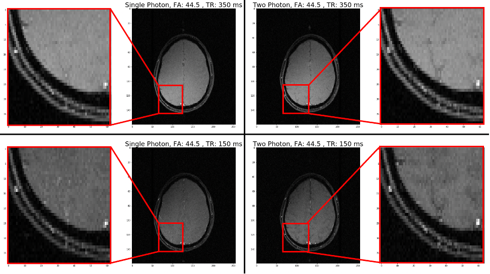
Figure 3: Single- and two-photon GRE brain images of a healthy volunteer. Left panel: Single-photon image with a flip angle of 44.5 degrees vs. a TR of 350 ms (Top) and a TR of 150 ms (Bottom). Right panel: Two-photon image with a $$$B_{1xy}$$$ amplitude corresponding to an equivalent two-photon 44.5-degree flip vs. a TR of 350 ms (Top) and a TR of 150 ms (Bottom). Some brain structure contrast is seen to be enhanced. Other parameters: FOV 27.6 cm, slice thickness 3 mm, matrix 256x160, TE 10ms.
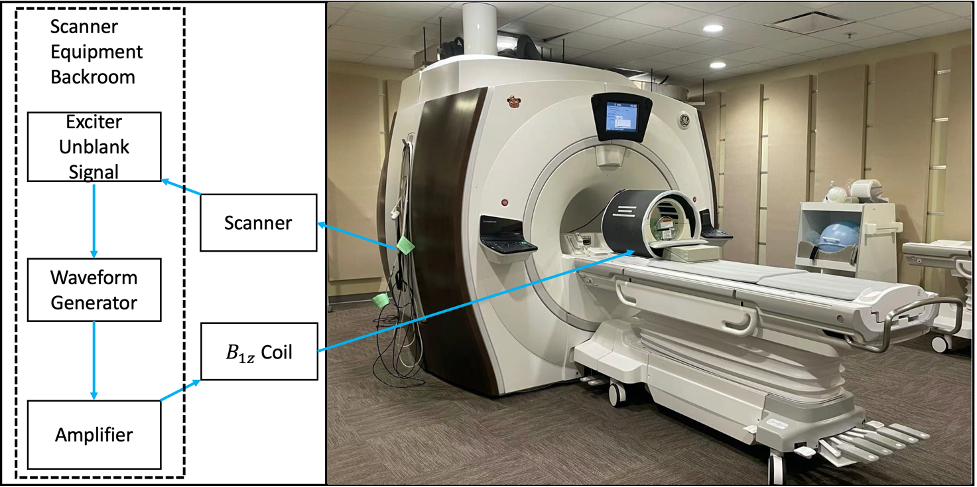
Figure 2: Left: equipment room set up. Right: $$$B_{1z}$$$ coil in a 3T scanner.