Michael Bernier1,2, Jingyuan E Chen1,2, Olivia Viessmann1,2, Nina E Fultz1,3, Maxime Chamberland4, Rebecca K Leaf5, Lawrence L Wald1,2,6, and Jonathan R Polimeni1,2,6
1Department of Radiology, A. A. Martinos Center for Biomedical Imaging, Massachusetts General Hospital, Boston, MA, United States, 2Radiology, Harvard Medical School, Boston, MA, United States, 3Department of Engineering, University of Boston, Boston, MA, United States, 4Cardiff University Brain Research Imaging Centre (CUBRIC), Cardiff, United Kingdom, 5Division of Hematology, Massachusetts General Hospital, Boston, MA, United States, 6Division of Health Sciences and Technology, Massachusetts Institute of Technology, Boston, MA, United States
1Department of Radiology, A. A. Martinos Center for Biomedical Imaging, Massachusetts General Hospital, Boston, MA, United States, 2Radiology, Harvard Medical School, Boston, MA, United States, 3Department of Engineering, University of Boston, Boston, MA, United States, 4Cardiff University Brain Research Imaging Centre (CUBRIC), Cardiff, United Kingdom, 5Division of Hematology, Massachusetts General Hospital, Boston, MA, United States, 6Division of Health Sciences and Technology, Massachusetts Institute of Technology, Boston, MA, United States
We used a blood-pool
contrast agent to increase four-fold the segmented vasculature, and developed an approach to reconstruct the vascular pathways by approximating spherical harmonics. We used deterministic tractography on the ODFs to compute a vascular connectome of GM and WM atlases.
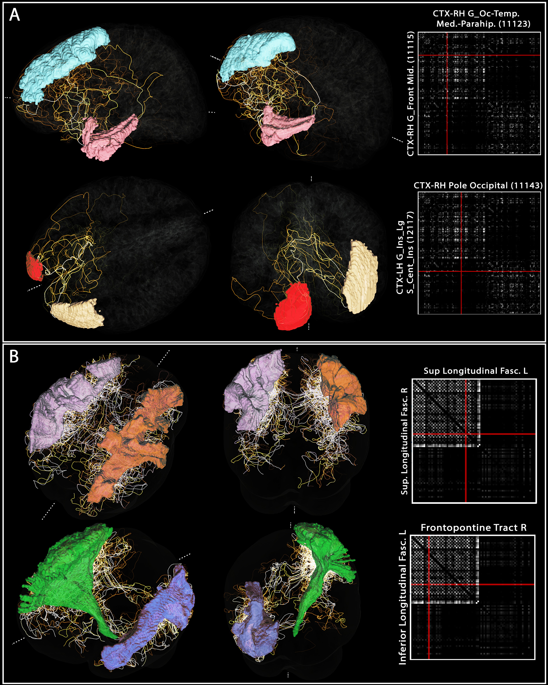
3. Example “vascular connectome” and specific
vascular pathways connecting pairs of brain regions. For both (A) Freesurfer’s Destrieux 2009 GM parcellation
and (B) Recobundle White-Matter bundle atlas, the connectome obtained for one
subject is shown. For each, two example region-to-region connections were
selected from the tracts computed previously to illustrate the complex pathway
connecting those regions.
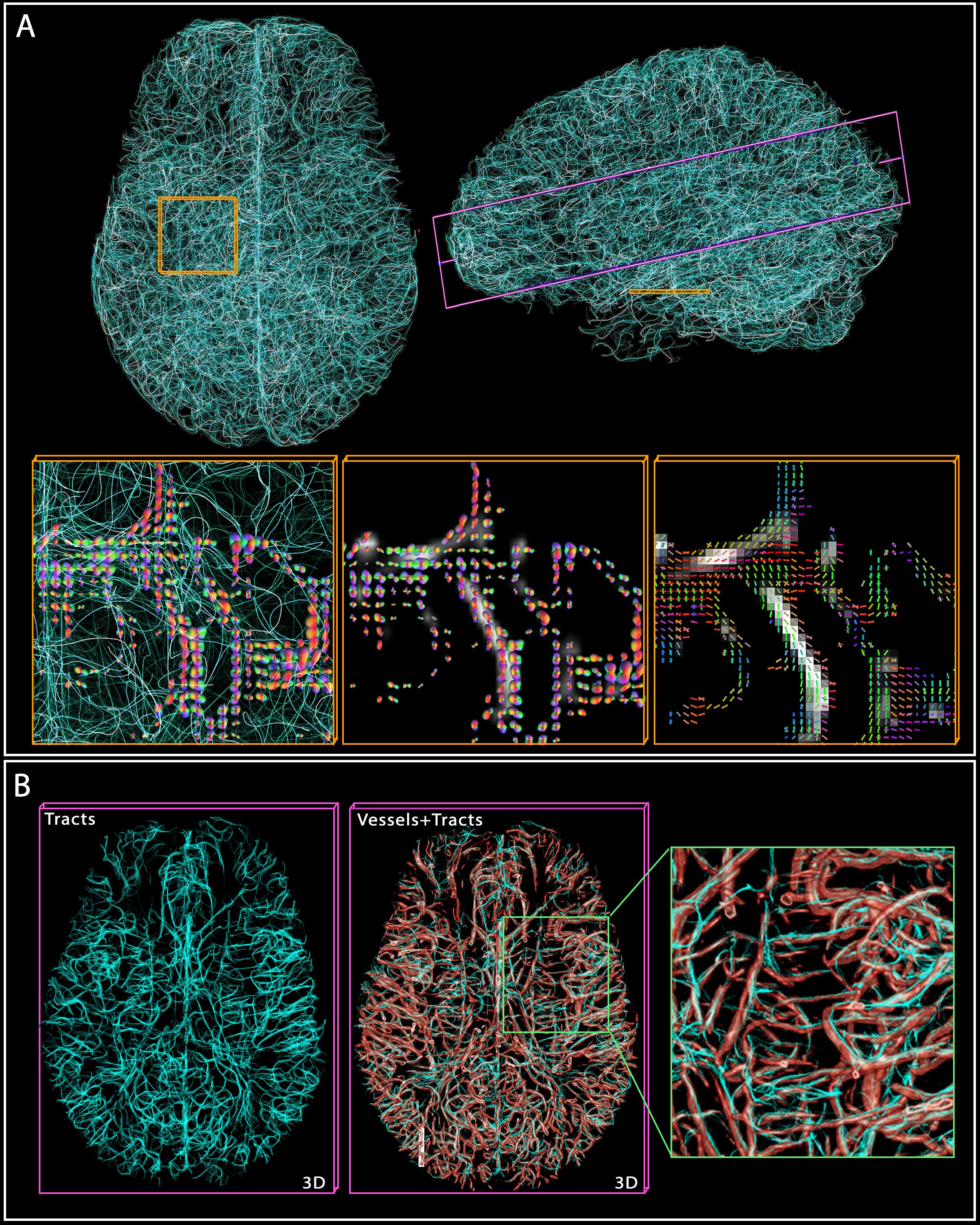
2. Vascular tractography. (A) shows 10k tracts reconstructed from the 200k computed, with two cross-sections
highlighted in yellow and purple. In yellow, the tracts and ‘vesselness’ scores
are overlaid by the ODF at crossings, while the rightmost shows the
main directions (tensor) of the ODFs. (B) In the purple cross-section, the
tracts and vessels are overlaid with transparency to demonstrate the
correspondence.