Kengo Takahashi1,2, Filip Sobczak1,2, Patricia Pais-Roldán3, and Xin Yu1,4
1High-field Magnetic Resonance, Max Planck Institute for Biological Cybernetics, Tübingen, Germany, 2Graduate Training Centre of Neuroscience, Tübingen, Germany, 3Institute of Neuroscience and Medicine (INM-4), Forschungszentrum Jülich, Jülich, Germany, 4Athinoula A. Martinos Center, Massachusetts General Hospital and Harvard Medical School, Boston, MA, United States
1High-field Magnetic Resonance, Max Planck Institute for Biological Cybernetics, Tübingen, Germany, 2Graduate Training Centre of Neuroscience, Tübingen, Germany, 3Institute of Neuroscience and Medicine (INM-4), Forschungszentrum Jülich, Jülich, Germany, 4Athinoula A. Martinos Center, Massachusetts General Hospital and Harvard Medical School, Boston, MA, United States
fMRI signals in the lateral hypothalamus
coupling together with pupil dynamics may represent different brain states.
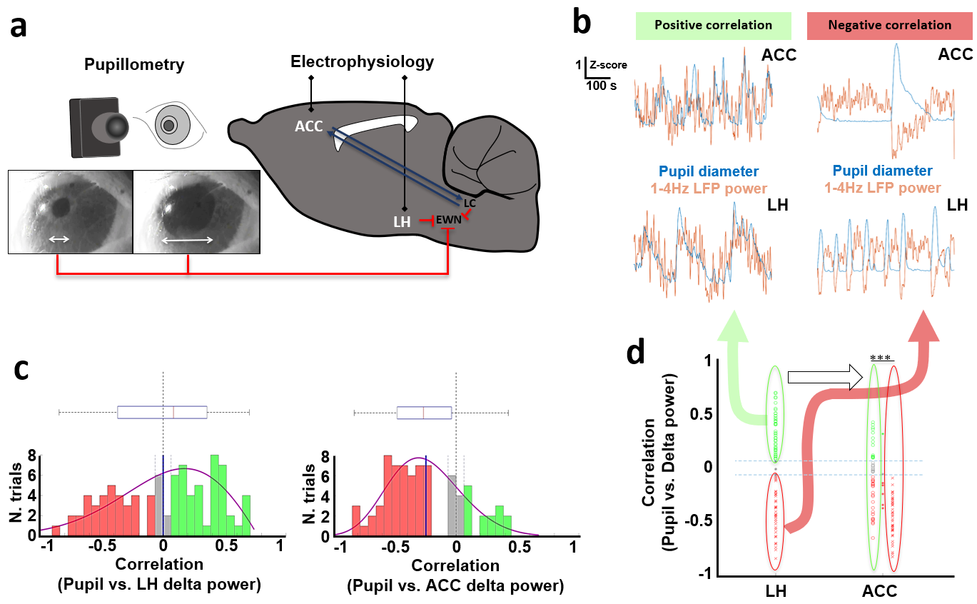
Figure 1. Electrophysiology
setup and correlations between pupil dynamics and LFP delta power from the ACC
or LH. a)
the experimental setup for
electrophysiology and pupil detection. b)
examples of positive and negative correlations between pupil diameter and LFP
delta (1-4Hz) power changes from the ACC and LH during 15-min resting state trials.
c) Distribution of correlation
coefficient between pupil dynamics and LFP delta band power at the LH or ACC. d) Categorization of trials based on a
positive or negative correlation between pupil dynamics and LH delta power.
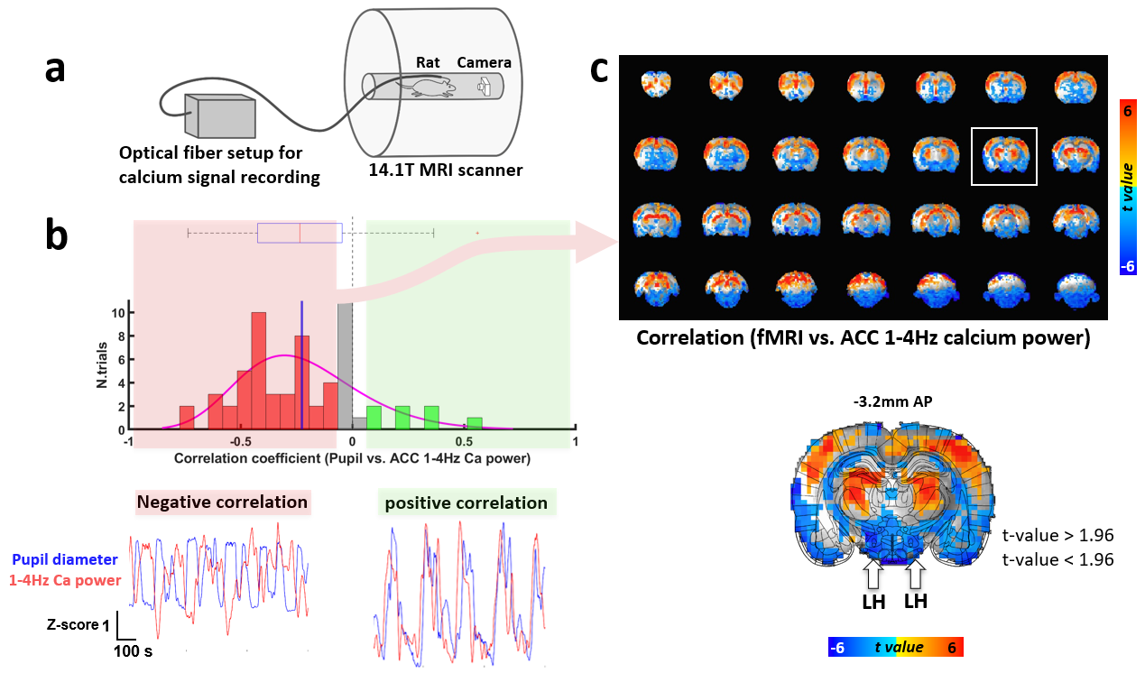
Figure 3. Correlations
between fMRI and ACC 1-4Hz calcium power in the whole brain. a) multimodal
resting-state fMRI setup simultaneously recorded with optical fiber calcium
signals and pupil dynamics. b) Distribution of correlation
coefficient between pupil dynamics and ACC 1-4Hz calcium power and their
examples of positive and negative correlations during 15-min trials. c) t-value maps of the correlation between fMRI and
ACC 1-4Hz calcium power when the pupil dynamics and ACC 1-4Hz calcium power
show negative correlations and location of the LH, indicated with white arrows.