Joana Cabral1,2, Francisca F. Fernandes1, and Noam Shemesh1
1Champalimaud Research, Champalimaud Centre for the Unknown, Lisbon, Portugal, 2Life and Health Sciences Research Institute, University of Minho, Braga, Portugal
1Champalimaud Research, Champalimaud Centre for the Unknown, Lisbon, Portugal, 2Life and Health Sciences Research Institute, University of Minho, Braga, Portugal
Resonant oscillations in ultra-fast fMRI signal (TR = 38 ms) are detected within the rat cortex under sedation, peaking between 0.03z and 0.25Hz and synchronizing in phase across distant brain areas, providing insights into the mechanisms underlying resting-state functional connectivity.
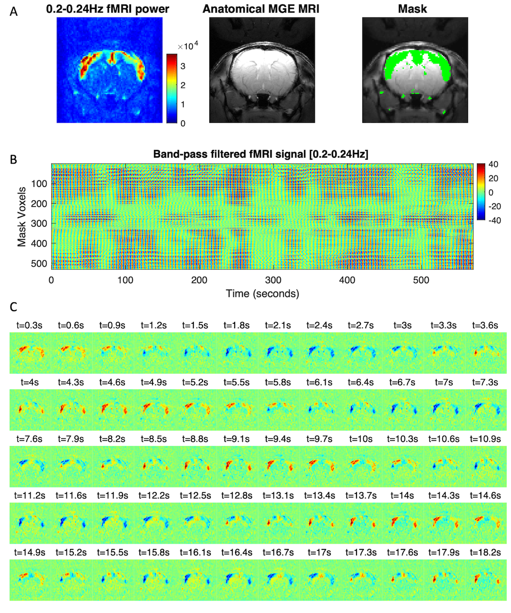
Bilaterally synchronized cortex-specific oscillations in fMRI signal detected at 0.20-0.24Hz. A – A mask is defined by considering all voxels exhibiting power 5 standard deviations above the mean level detected in a dead rat. B – The fMRI signal in the mask voxels is band-pass filtered between 0.20 and 0.24 Hz and plotted over time, revealing ultra-slow amplitude modulations (here one representative scan). C – Snap shots of the band-pass filtered fMRI signal revealing bilateral synchronization.
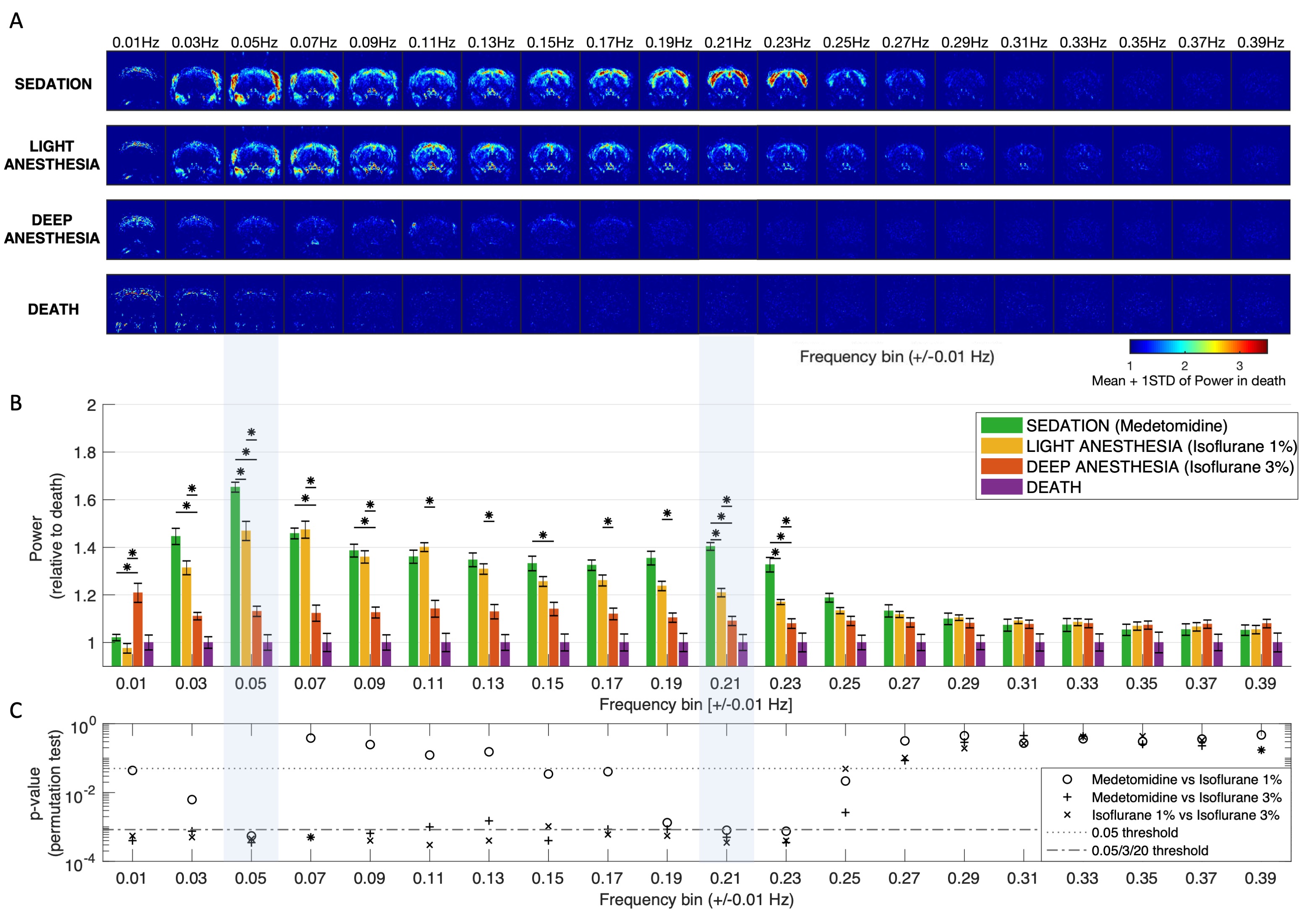
Slow oscillations decrease in power with anesthesia. A – fMRI Spectral power maps (averaged across scans) reveal resonance at specific frequencies in different brain and body structures that decrease significantly in power under deep anesthesia. B – Comparison of the power in each frequency bin between conditions (6 scans per condition), normalized by the average power in the dead rat scans. C – Statistically significant differences are only considered if p<0.00083 (Bonferroni threshold).