Benjamin Marty1,2, Fabian Balsiger3, Pierre-Yves Baudin1,2, Lopez Alfredo1,2, Ericky CA Araujo1,2, and Harmen Reyngoudt1,2
1Neuromuscular Investigation Center, NMR Laboratory, Institute of Myology, Paris, France, 2NMR Laboratory, CEA, DRF, IBFJ, MIRCen, Paris, France, 3Support Center for Advanced Neuroimaging, Institute for Diagnostic and Interventional Neuroradiology, Inselspital, Bern University Hospital, University of Bern, Bern, Switzerland
1Neuromuscular Investigation Center, NMR Laboratory, Institute of Myology, Paris, France, 2NMR Laboratory, CEA, DRF, IBFJ, MIRCen, Paris, France, 3Support Center for Advanced Neuroimaging, Institute for Diagnostic and Interventional Neuroradiology, Inselspital, Bern University Hospital, University of Bern, Bern, Switzerland
We determined the influence of the reconstruction
parameters on fat fraction and water T1 estimated in the skeletal muscle
using MR fingerprinting with water and fat separation and proposed an
optimized reconstruction procedure maximizing the accuracy and precision
of the
MRI variables
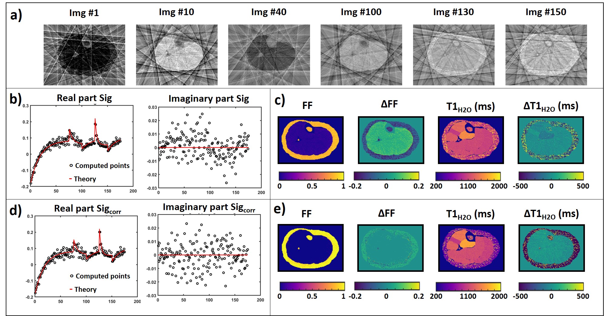
Figure 2: a)
Image series from numerical leg #1 using 8 spokes
per frame. b) Computed (black circles) and theoretical (red lines) real and
imaginary parts of the signal evolution measured in a muscle ROI. c)
FF and T1H2O maps, and difference maps
(versus the theoretical maps) obtained without correction of the
aliasing artifacts. d) Computed (black circles) and theoretical (red lines)
real and imaginary parts of the corrected signal evolution measured in the same ROI. e) FF and T1H2O maps, and difference maps obtained on leg #1 with correction of the aliasing artifacts.
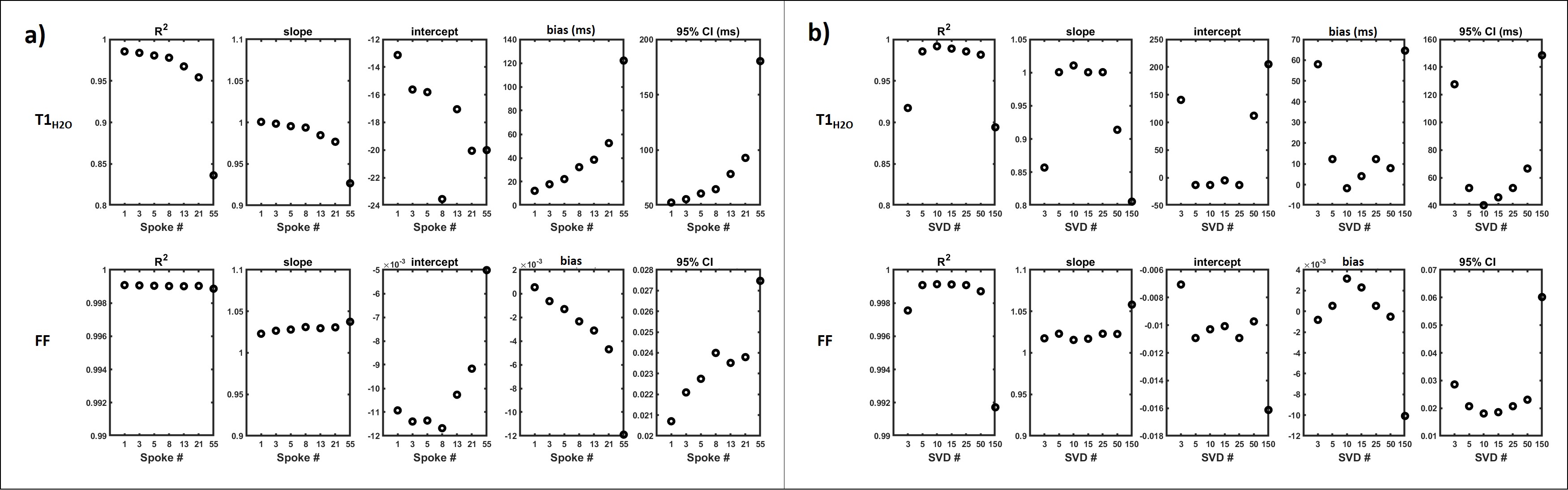
Figure 3: a) Influence of the number of spokes used
for reconstructing the MRF T1-FF image series on the Pearson correlation
coefficient (r2), slope and
intercept of the linear correlation, bias and 95% confidence interval (CI)
between the computed and the theoretical FF and T1H2O values. b)
Influence of the number of SVD components used for dictionary matching on the
Pearson correlation coefficient (r2),
slope and intercept of the linear correlation, bias and 95% confidence interval
(CI) between the computed and the theoretical FF and T1H2O values.