Sophia Swago1, Abigail Cember2, Brianna Moon1, Puneet Bagga3, Neil Wilson4, Mark A. Elliott2, Hari Hariharan2, Ravinder Reddy2, and Walter Witschey2
1Department of Bioengineering, University of Pennsylvania, Philadelphia, PA, United States, 2Department of Radiology, University of Pennsylvania, Philadelphia, PA, United States, 3St. Jude Children's Research Hospital, Memphis, TN, United States, 4Siemens Medical Solutions USA Inc, Malven, PA, United States
1Department of Bioengineering, University of Pennsylvania, Philadelphia, PA, United States, 2Department of Radiology, University of Pennsylvania, Philadelphia, PA, United States, 3St. Jude Children's Research Hospital, Memphis, TN, United States, 4Siemens Medical Solutions USA Inc, Malven, PA, United States
To characterize cross-relaxation properties of downfield 1H resonances
in skeletal muscle in vivo, we compare the T1 of three resonances with
selective and nonselective inversion. The latter increases the T1 of the resonances
at 8.0, 8.2 and 8.5 ppm.
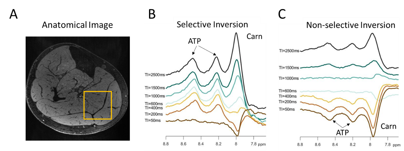
Figure 2: Single-voxel downfield spectra. A) Position
of the voxel from which spectra are collected. Representative downfield spectra
under selective (B) and non-selective inversion (C) conditions show potential
ATP resonances at 8.2 and 8.5 ppm, and a carnosine resonance at 7.9-8.0 ppm.
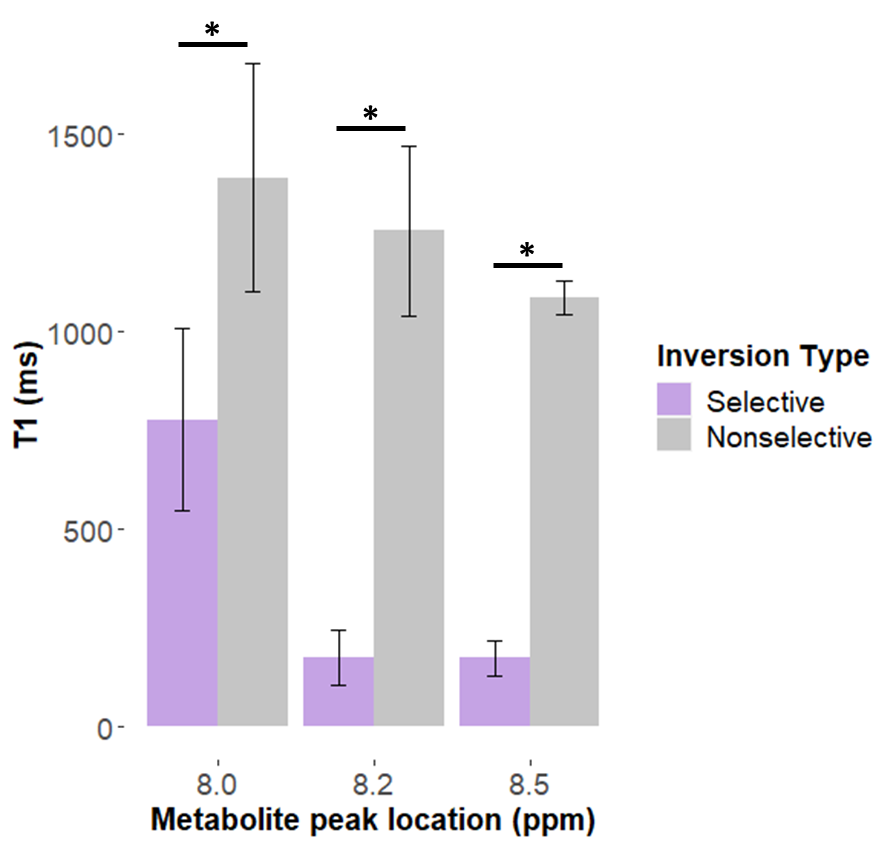
Figure 4: Apparent T1 of downfield resonances after
selective and nonselective inversion. For each resonance, the T1 under
nonselective inversion was significantly longer than under selective inversion.
* indicates p<0.01.