Laura Saunders1, Paul J. C. Hughes1, Dave Capener1, David G Kiely1,2, Jim M Wild1, and Andy J Swift1
1Infection Immunity and Cardiovascular Disease, University of Sheffield, Sheffield, United Kingdom, 2Sheffield Pulmonary Vascular Disease Unit, Sheffield, United Kingdom
1Infection Immunity and Cardiovascular Disease, University of Sheffield, Sheffield, United Kingdom, 2Sheffield Pulmonary Vascular Disease Unit, Sheffield, United Kingdom
Lung M0 maps may allow differentiation of perfusion defects in patients
with CTEPH/CTED from other patients. Patients with CTEPH/CTED had lower M0 in non-perfused
lung, whereas control patients did not. Lung T1 was significantly lower in perfusion defects in all
patients.
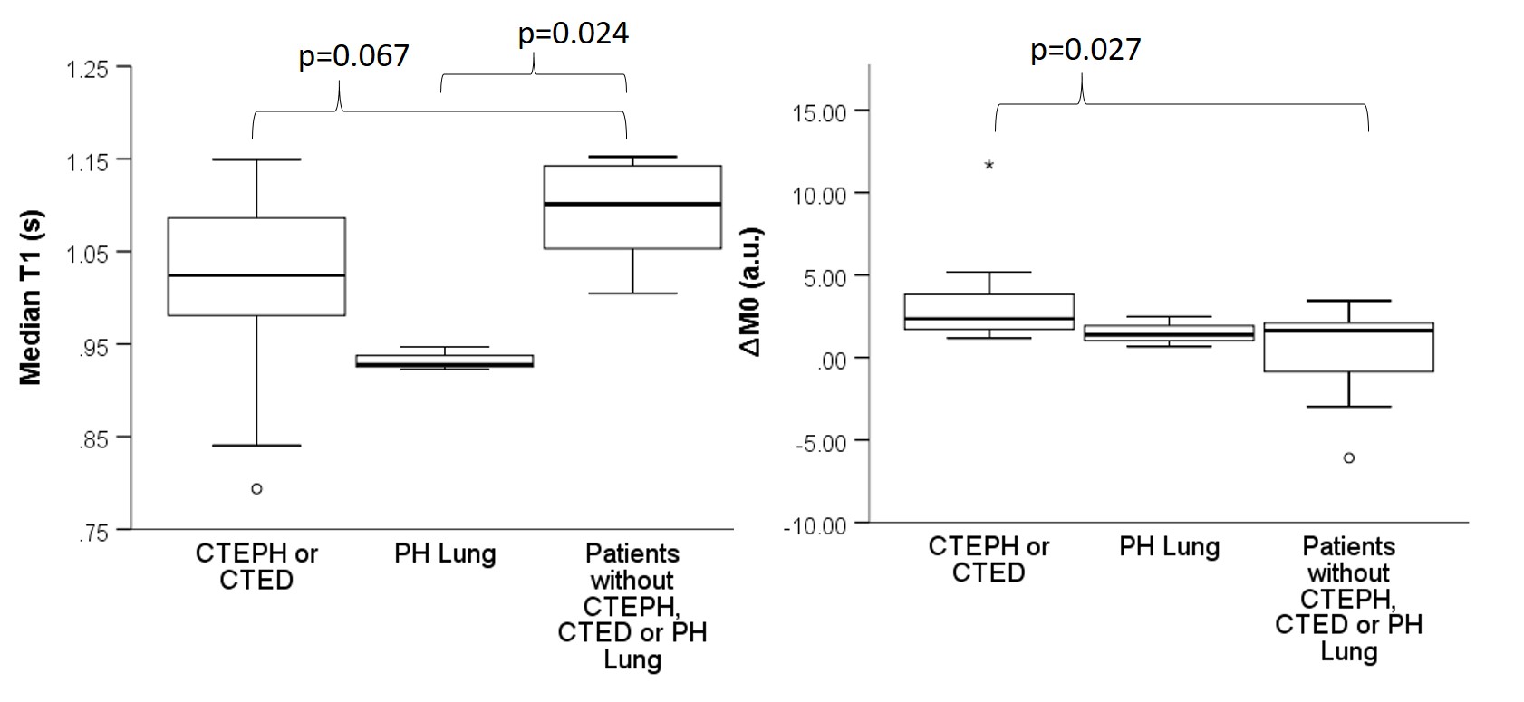
Figure 4: Median T1 was significantly lower
in patients with CTEPH/CTED and PH-lung compared to controls. ∆M0 was significantly higher
in patients with CTEPH/CTED than controls.
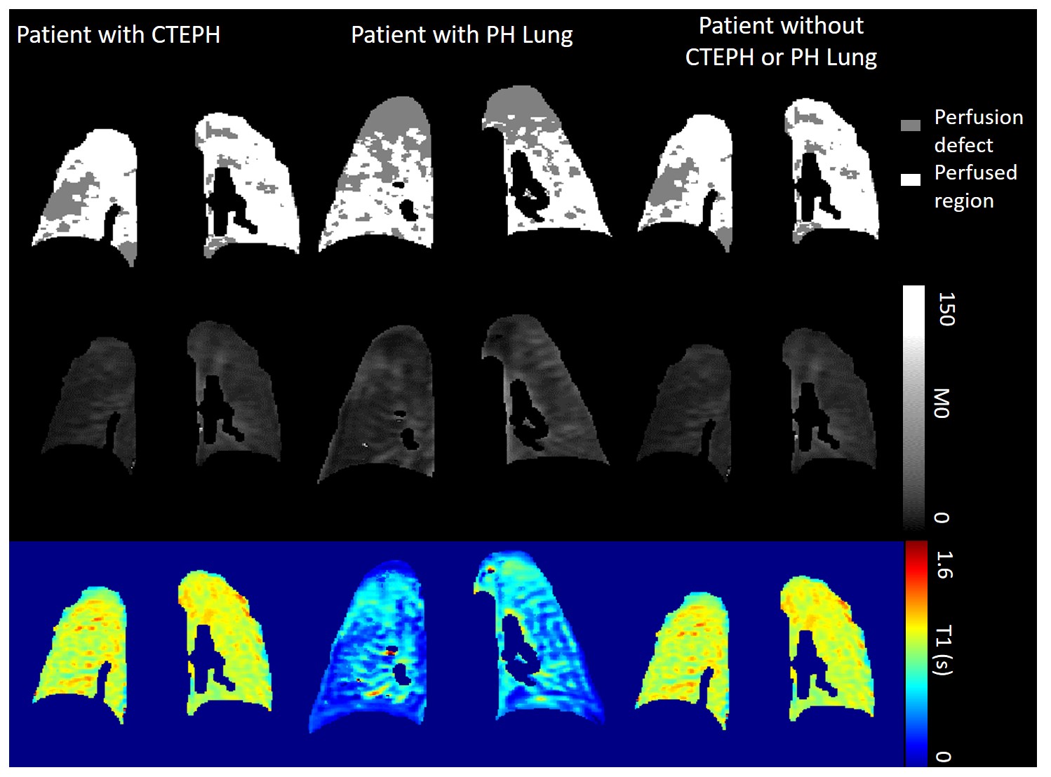
Figure
2: Segmentations of perfused and non-perfused regions were applied to M0 and T1
maps, to calculate median M0 and T1 in these regions. Example maps
are shown for a patient with CTEPH, a patient with PH lung and a control
patient.