1Radiology, Fujita Health University School of Medicine, Toyoake, Japan, 2Joint Research Laboratory of Advanced Medical Imaging, Fujita Health University School of Medicine, Toyoake, Japan, 3Division of Functional and Diagnostic Imaging Research, Department of Radiology, Kobe University Graduate School of Medicine, Kobe, Japan, 4Canon Medical Systems Corporation, Otawara, Japan, 5Diagnostic Radiology, Hyogo Cancer Center, Akashi, Japan
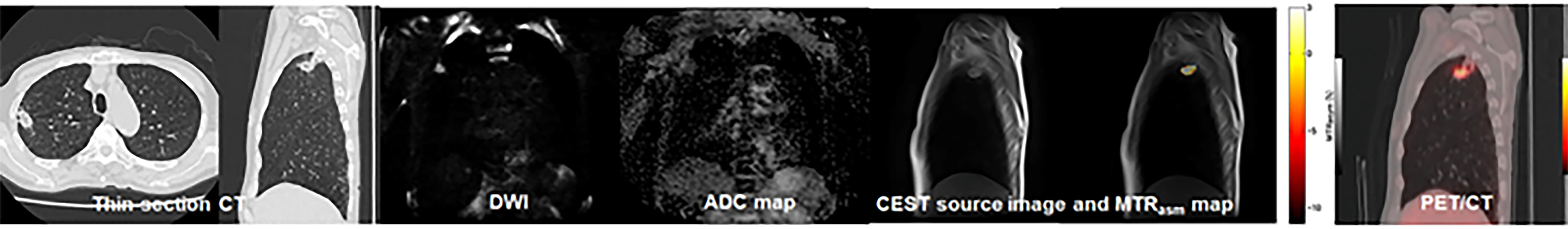
Figure 1. 62-year-old female with invasive adenocarcinoma (L to R: thin-section CT, DWI, ADC map, MTRasym map fused with T2WI, and SUVmax map fused with CT) and determined as recurrence group.
Thin-section CT demonstrates a nodule in the right upper lobe. ADC of this nodule was 1.05×10⁻³mm2/s. SUVmax was 2.6. APTw image shows low MTRasym with the value of 1.25. This case was false-negative on PET/CT, and true-positive on DWI and APTw image. When combined both indexes, PET/CT with APTw image was diagnosed as recurrence group and determined as true-positive case.
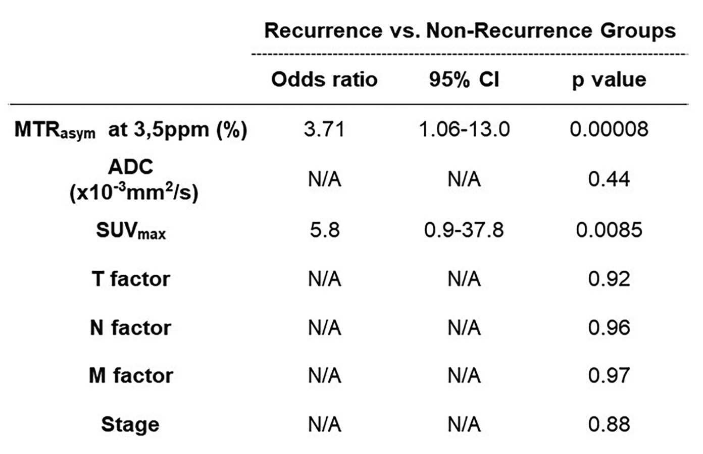
Figure 2. Results of multivariate regression analysis for distinguishing recurrence from non-recurrence groups in NSCLC patients.
MTRasym at 3.5ppm and SUVmax were determined as significant predictor for distinguishing recurrence from non-recurrence groups (p<0.05).