Na Hu1, Tianwei Zhang2, Yifan Wu3, Biqiu Tang1, Minlong Li1, Qiyong Gong1, Shi Gu2, and Su Lui1
1Huaxi MR Research Center (HMRRC), Department of Radiology, West China Hospital of Sichuan University, Chengdu, China, 2Department of Computer and Engineering, University of Electronic Science and Technology of China, Chengdu, China, 3Department of Bioengineering, University of Pennsylvania, Philadelphia, PA, United States
1Huaxi MR Research Center (HMRRC), Department of Radiology, West China Hospital of Sichuan University, Chengdu, China, 2Department of Computer and Engineering, University of Electronic Science and Technology of China, Chengdu, China, 3Department of Bioengineering, University of Pennsylvania, Philadelphia, PA, United States
With synthetic MRI compared to CT, sensitivity was
improved by 116% in patient detection and 300% in lesion detection, and extra 75%
of patients and 15% of lesions missed on CT were detected on synthetic MRI.
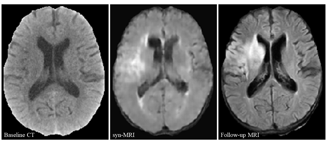
Figure 3. Example of patient detection using synthetic MRI (syn-MRI) versus CT in the testing
set.
Brain baseline CT (left) fails to show any definite hypoattenuating
lesions, although the gray-white matter junction of the right insula is
suspected. Synthetic MRI (middle) shows distinct hyperintensity in the
territory of middle cerebral artery on the right, which is corresponding to the
finding on the follow-up MRI (right).
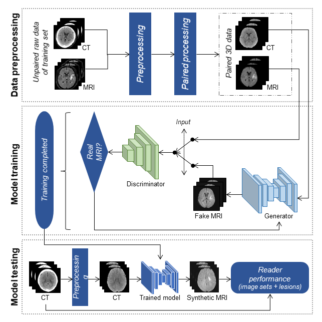
Figure 1. Training and
Testing of the generative adversarial network model.