Po-Jui Lu1,2,3, Muhamed Barakovic1,2,3, Matthias Weigel1,2,3,4, Reza Rahmanzadeh1,2,3, Riccardo Galbusera1,2,3, Simona Schiavi5, Alessandro Daducci5, Francesco La Rosa6,7,8, Meritxell Bach Cuadra6,7,8, Robin Sandkühler9, Jens Kuhle2,3, Ludwig Kappos2,3, Philippe Cattin9, and Cristina Granziera1,2,3
1Translational Imaging in Neurology (ThINk) Basel, Department of Biomedical Engineering, University Hospital Basel and University of Basel, Basel, Switzerland, 2Neurology Clinic and Policlinic, Departments of Medicine, Clinical Research and Biomedical Engineering, University Hospital Basel and University of Basel, Basel, Switzerland, 3Research Center for Clinical Neuroimmunology and Neuroscience (RC2NB) Basel, University Hospital Basel and University of Basel, Basel, Switzerland, 4Division of Radiological Physics, Department of Radiology, University Hospital Basel, Basel, Switzerland, 5Department of Computer Science, University of Verona, Verona, Italy, 6Signal Processing Laboratory (LTS5), Ecole Polytechnique Fédérale de Lausanne, Lausanne, Switzerland, 7Medical Image Analysis Laboratory, Center for Biomedical Imaging (CIBM), University of Lausanne, Lausanne, Switzerland, 8Department of Radiology, Lausanne University Hospital and University of Lausanne, Lausanne, Switzerland, 9Center for medical Image Analysis & Navigation, Department of Biomedical Engineering, University of Basel, Allschwil, Switzerland
1Translational Imaging in Neurology (ThINk) Basel, Department of Biomedical Engineering, University Hospital Basel and University of Basel, Basel, Switzerland, 2Neurology Clinic and Policlinic, Departments of Medicine, Clinical Research and Biomedical Engineering, University Hospital Basel and University of Basel, Basel, Switzerland, 3Research Center for Clinical Neuroimmunology and Neuroscience (RC2NB) Basel, University Hospital Basel and University of Basel, Basel, Switzerland, 4Division of Radiological Physics, Department of Radiology, University Hospital Basel, Basel, Switzerland, 5Department of Computer Science, University of Verona, Verona, Italy, 6Signal Processing Laboratory (LTS5), Ecole Polytechnique Fédérale de Lausanne, Lausanne, Switzerland, 7Medical Image Analysis Laboratory, Center for Biomedical Imaging (CIBM), University of Lausanne, Lausanne, Switzerland, 8Department of Radiology, Lausanne University Hospital and University of Lausanne, Lausanne, Switzerland, 9Center for medical Image Analysis & Navigation, Department of Biomedical Engineering, University of Basel, Allschwil, Switzerland
GAMER MRI can select
discriminating diffusion measures from diffusion models in the
classification of multiple sclerosis lesions. The combinations of selected
measures have strong correlation with the Expanded Disability Status Scale and
the serum level of neurofilament light chain.
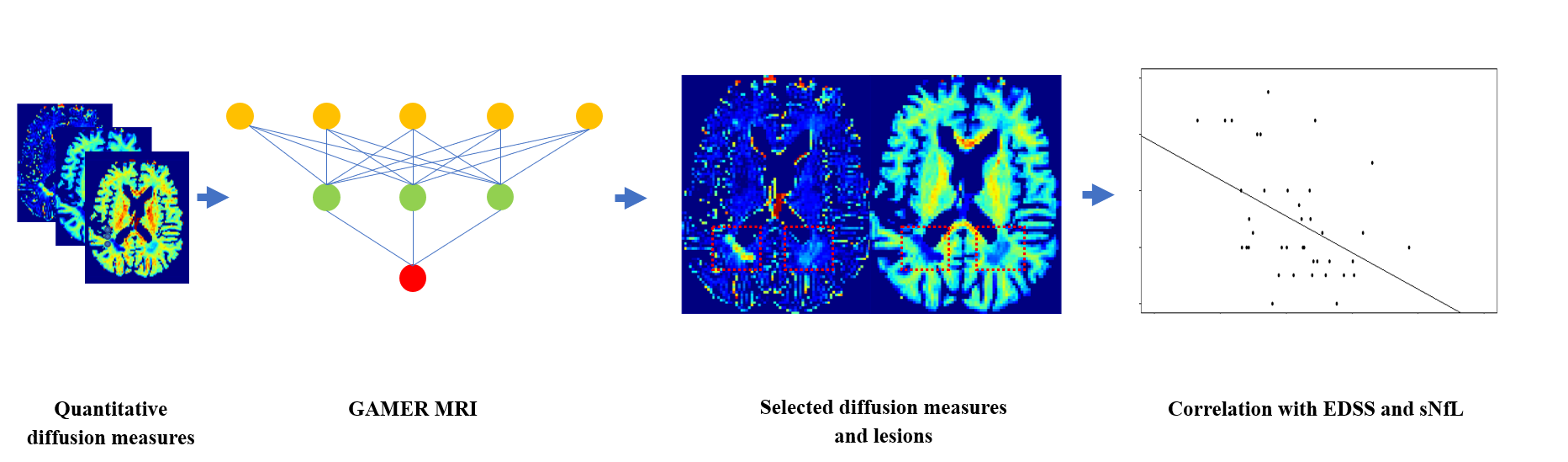
Fig. 1: Flowchart for using GAMER-MRI to
select the most discriminating subject-wise normalized diffusion measures and correlating the combinations of selected diffusion measures with the
Expanded Disability Status Scale and the serum level of neurofilament light
chain.
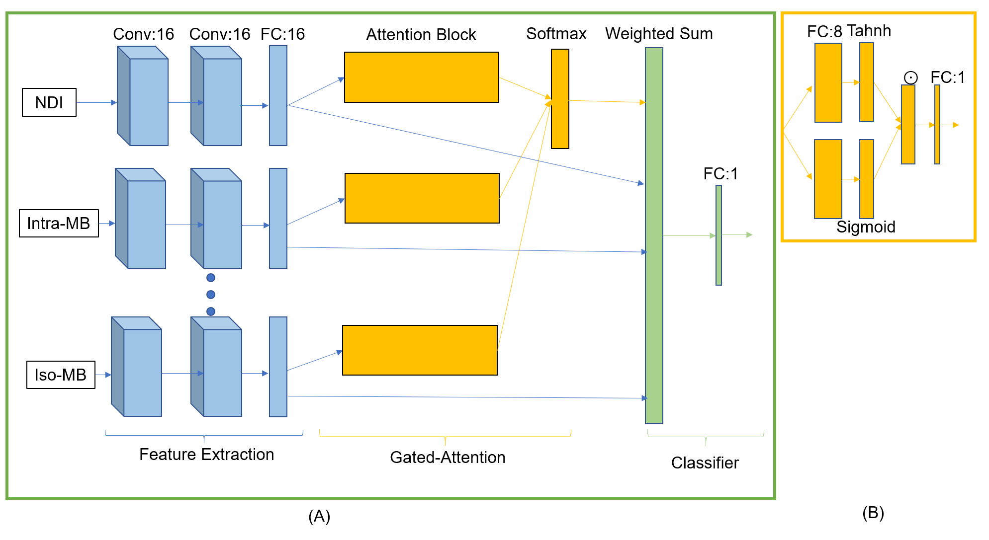
Fig. 2: GAMER-MRI. (A) The neural
network. Conv is a convolutional block consisting of a 3x3x3 convolutional
layer, exponential leaky units and batch normalization. FC is a fully connected
layer. Attention weights are obtained
from the softmax function after the attention blocks. Each diffusion measure is
encoded parallelly before the softmax function. The hidden features of
diffusion measures are linearly combined with the attention weights and input
to the classifier. (B) Attention block. ⊙ represents an element-wise multiplication.