Nam G. Lee1, Rajiv Ramasawmy2, Adrienne E. Campbell-Washburn2, and Krishna S. Nayak1,3
1Biomedical Engineering, University of Southern California, Los Angeles, CA, United States, 2Cardiovascular Branch, Division of Intramural Research, National Heart, Lung, and Blood Institute, National Institutes of Health, Bethesda, MD, United States, 3Ming Hsieh Department of Electrical and Computer Engineering, University of Southern California, Los Angeles, CA, United States
1Biomedical Engineering, University of Southern California, Los Angeles, CA, United States, 2Cardiovascular Branch, Division of Intramural Research, National Heart, Lung, and Blood Institute, National Institutes of Health, Bethesda, MD, United States, 3Ming Hsieh Department of Electrical and Computer Engineering, University of Southern California, Los Angeles, CA, United States
We present MaxGIRF, an image reconstruction
method to simultaneously mitigate local blurring caused by concomitant fields and
static off-resonance. We demonstrate successful application to 2D spin-echo
spiral brain imaging at 0.55T with (long) 12ms readouts.
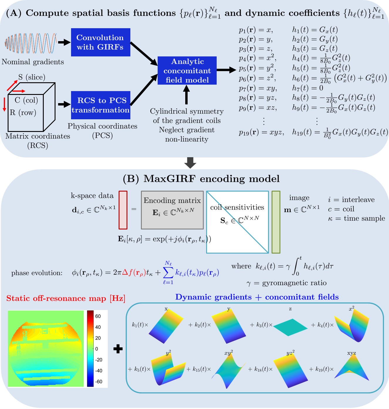
Figure 1. Flowchart of the
proposed MaxGIRF reconstruction. (A) Logical gradient waveforms are first transformed to the physical coordinate system (PCS). Gradient inaccuracies in PCS are estimated using phantom-based
GIRFs. Analytic expressions of concomitant fields are then
derived from the coil geometry, presumed gradient non-linearity, and GIRF-predicted
gradients, for each spatial position in PCS. (B) The phase evolution per voxel
is represented as the sum of phase contributions from static off-resonance
(red) and linear gradients and
concomitant field terms (blue).
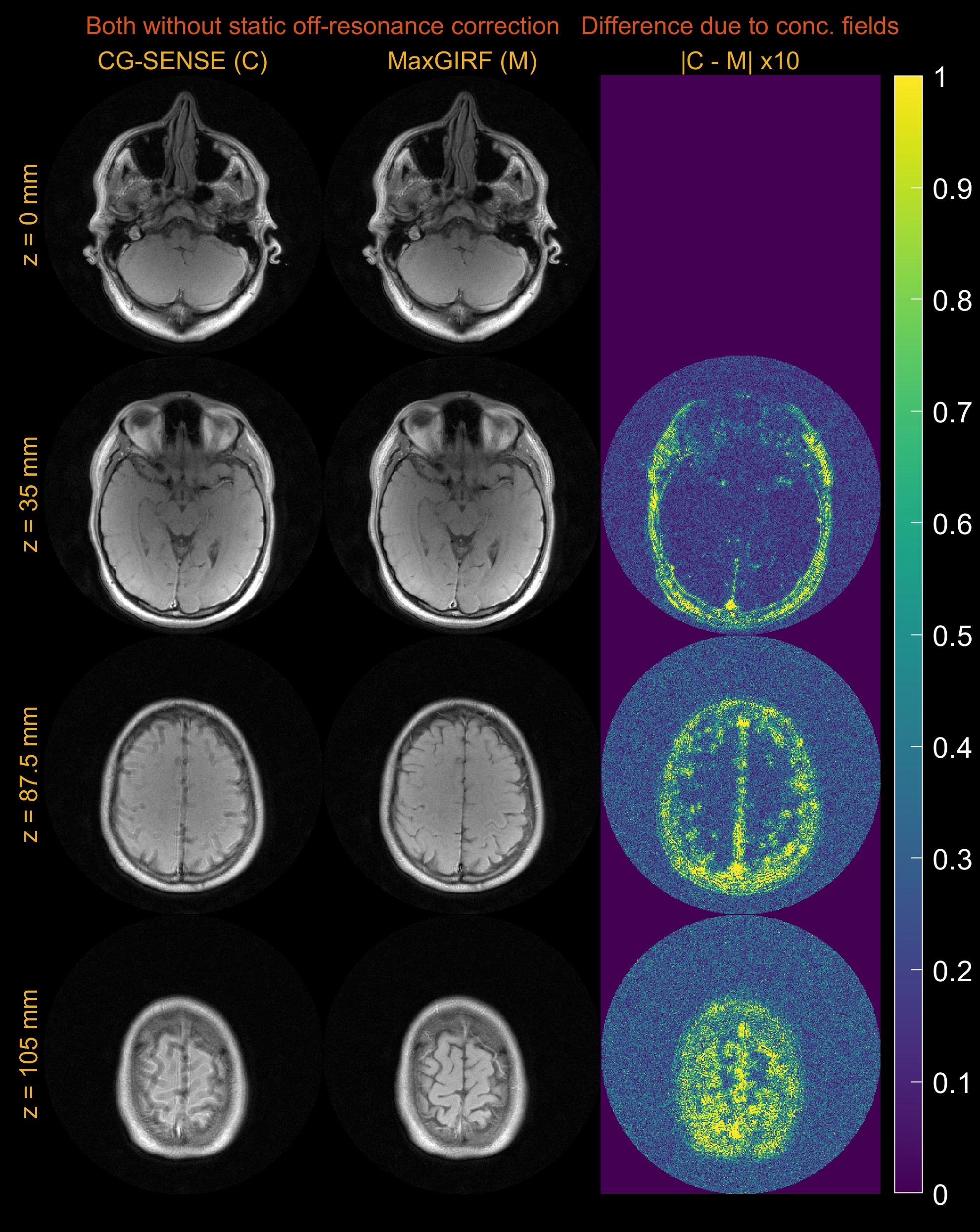
Figure 4. Multi-slice axial spiral imaging of a healthy volunteer at 0.55T. (left) CG-SENSE reconstructions; (middle) MaxGIRF reconstructions without static off-resonance correction (i.e., without a field map); (right) Absolute difference images. GIRF-predicted gradients were used in bothreconstructions. The improvement in concomitant field blurring is evident in away from isocenter, as expected. Static off-resonance correction was not performed, in order to isolate the difference due to concomitant field correction.