Christian R. Meixner1, Sebastian Schmitter2,3, Jürgen Herrler4, Arnd Dörfler4, Michael Uder1, and Armin M. Nagel1,3
1Institute of Radiology, University Hospital Erlangen, Friedrich-Alexander-Universität Erlangen-Nürnberg, Erlangen, Germany, 2Physikalisch-Technische Bundesanstalt (PTB), Braunschweig und Berlin, Germany, 3Division of Medical Physics in Radiology, German Cancer Research Center (DKFZ), Heidelberg, Germany, 4Institute of Neuro-Radiology, Friedrich-Alexander Universität Erlangen-Nürnberg, Erlangen, Germany
1Institute of Radiology, University Hospital Erlangen, Friedrich-Alexander-Universität Erlangen-Nürnberg, Erlangen, Germany, 2Physikalisch-Technische Bundesanstalt (PTB), Braunschweig und Berlin, Germany, 3Division of Medical Physics in Radiology, German Cancer Research Center (DKFZ), Heidelberg, Germany, 4Institute of Neuro-Radiology, Friedrich-Alexander Universität Erlangen-Nürnberg, Erlangen, Germany
The combination of a B1+-shim
for the labeling and 2-spoke dynamic transmission pulses for the readout
improves 4D-pcASL angiography at 7T compared to the standard circular polarized
mode by an increased vessel intensity and more vessel conspicuity.

Figure 1: a) Hybrid
B1+ shimming for the labeling: the left image shows a MIP
of the TOF to obtain two regions of interest around the main feeding arteries.
The image in the middle shows the un-shimmed B1+ -map in
the CP mode and on the right the B1+ -map after B1+
shimming (at 82V, reference voltage = 269V). b) The 2-spoke pTx for the readout
(Bloch simulated flip angle maps): left: flip angle map in the CP-mode; right:
flip angle map with the 2-spoke pTx after the MLS optimization.
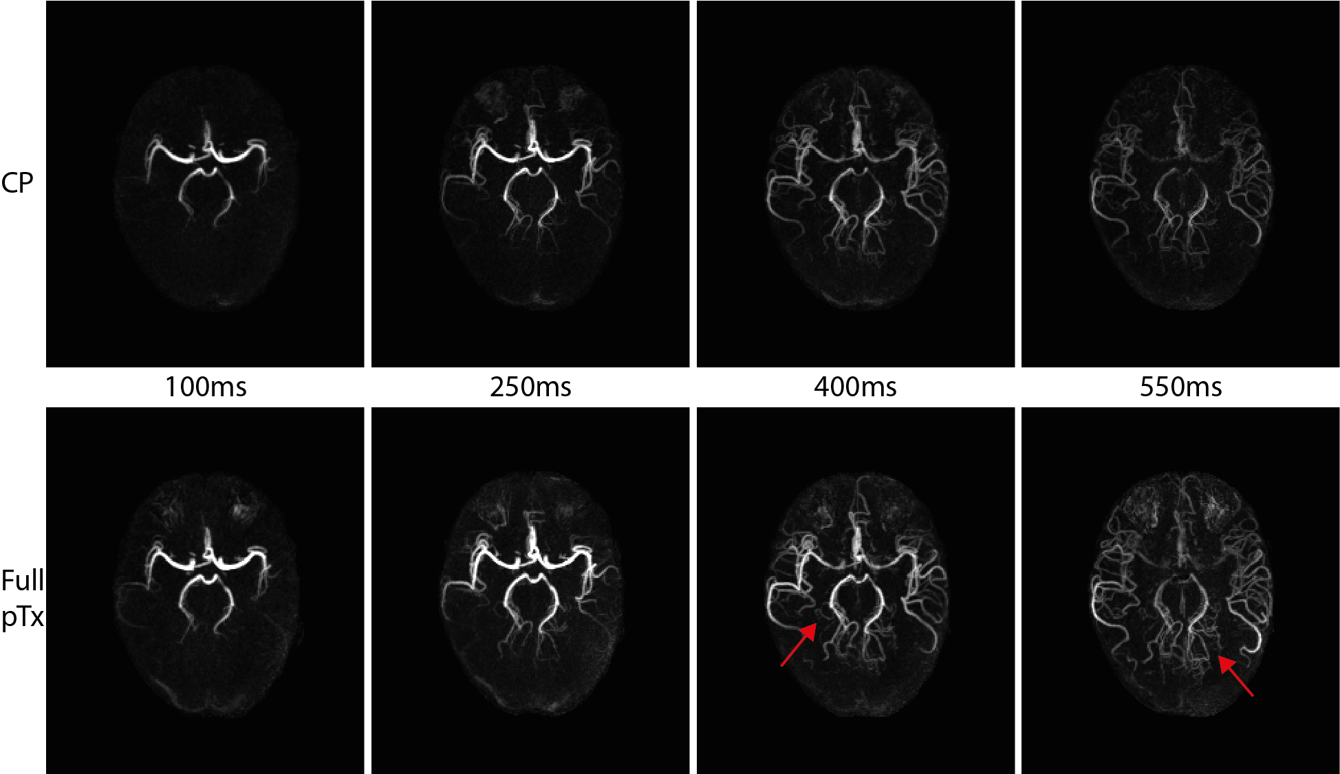
Figure 4:
Example of one subject in CP-mode (upper row) and with full pTx (lower row).
All images show the same windowing. It can be seen, that the signal intensity
in the vessels is higher with full pTx compared to CP-mode. The red arrows also
mark vessels which are not visible in the CP-mode but with full pTx.