Yuhui Chai1, Tina Liu1, Sean Marrett1, Linqing Li1, Arman Khojandi1, Daniel Handwerker1, Arjen Alink2, Lars Muckli3, and Peter Bandettini1
1NIMH, Bethesda, MD, United States, 2University Medical Centre Hamburg-Eppendorf, Hamburg, Germany, 3University of Glasgow, Glasgow, United Kingdom
1NIMH, Bethesda, MD, United States, 2University Medical Centre Hamburg-Eppendorf, Hamburg, Germany, 3University of Glasgow, Glasgow, United Kingdom
We report a division of auditory and visual processing in human anterior and posterior planum temporale, with each receiving feedback inputs with distinct mechanisms.
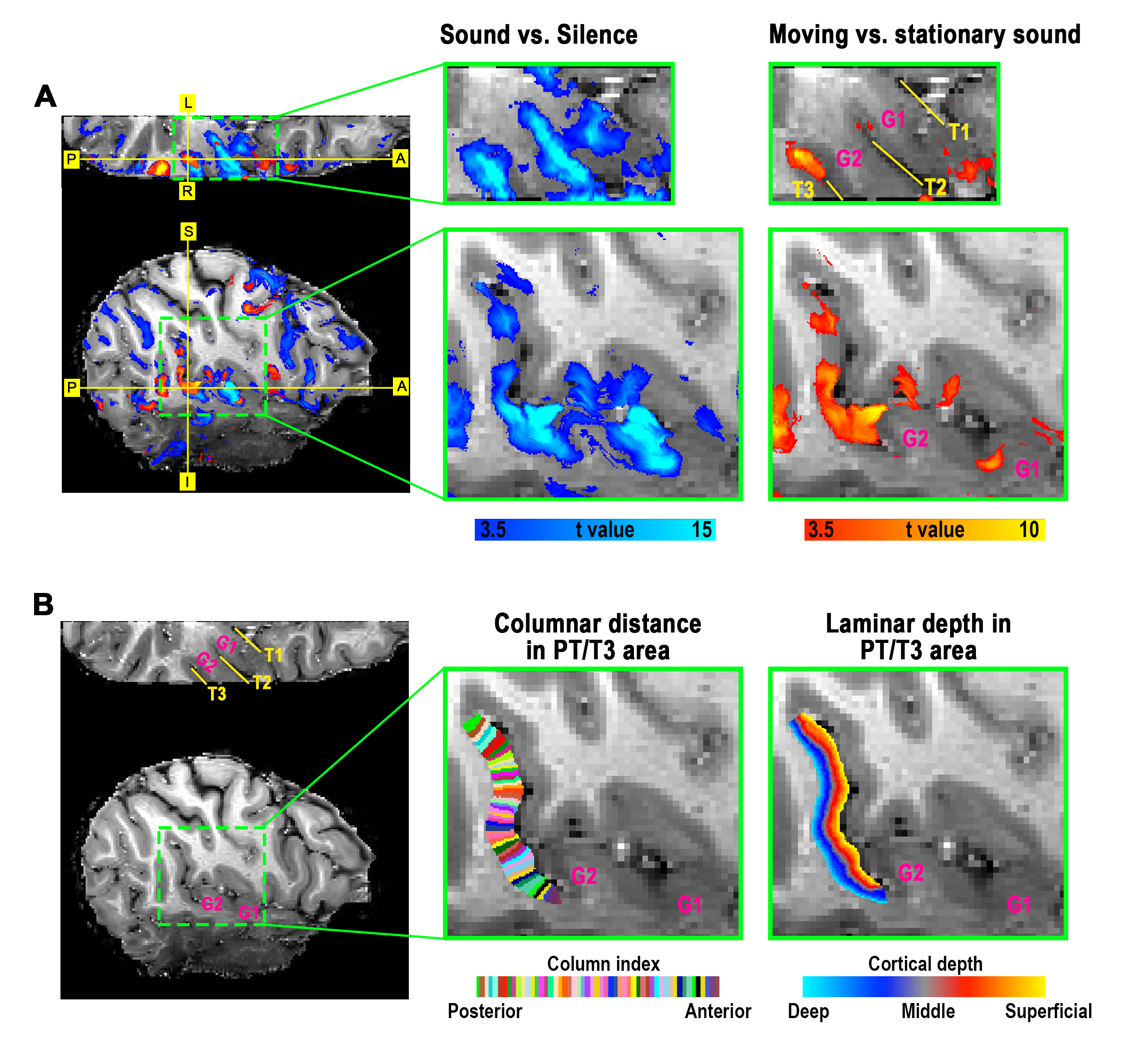
Figure 1. Localization of PT and its coordinate system across cortical columns and layers in one representative participant. All underlays show the anatomical EPI images which were obtained using an identical acquisition with functional images. (A) Overlays in blue and red show maps of BOLD signal changes induced by general sound (sound vs. silence) and movement-specific sound (moving sound vs. stationary sound), respectively. (B) Columnar distance and laminar depth determined in PT. G1 and G2 refer to the first and second transverse gyrus in auditory cortex, respectively.
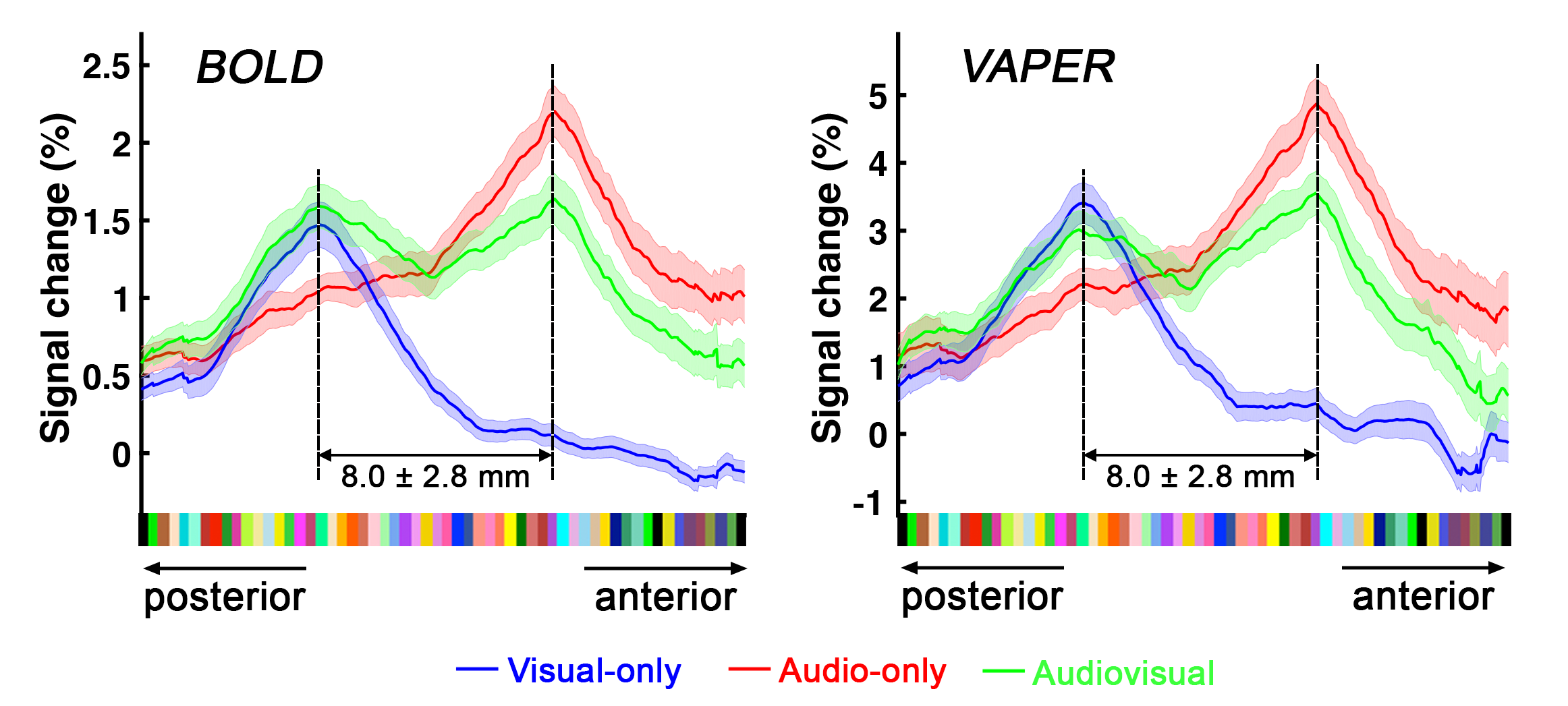
Figure 3. Group-averaged columnar profiles of sensory-specific representations under BOLD and VAPER contrasts. Blue, red and green curves represent signal changes in BOLD (left) and VAPER (right) to visual-only, audio-only and audiovisual stimuli, respectively. The distance between peaks of auditory and visual representations is 8 ± 2.8 mm along the cortical curvature. Error bars in the line plots represent ± SEM.