SoHyun Han1,2, HyungJoon Cho3, Kâmil Uludaǧ1,2, and Seong-Gi Kim1,2
1Center for Neuroscience Imaging Research, Suwon, Korea, Republic of, 2Department of Biomedical Engineering, Sungkyunkwan University, Suwon, Korea, Republic of, 3Department of Biomedical Engineering, Ulsan National Institute of Science and Technology, Ulsan, Korea, Republic of
1Center for Neuroscience Imaging Research, Suwon, Korea, Republic of, 2Department of Biomedical Engineering, Sungkyunkwan University, Suwon, Korea, Republic of, 3Department of Biomedical Engineering, Ulsan National Institute of Science and Technology, Ulsan, Korea, Republic of
Double SE-EPI
sequence was developed to achieve better sensitivity in SE-preparation and
demonstrated the feasibility of fMRI with 0.8-mm in-plane resolution, which can
be useful tool to layer-specific studies in humans with high specificity.
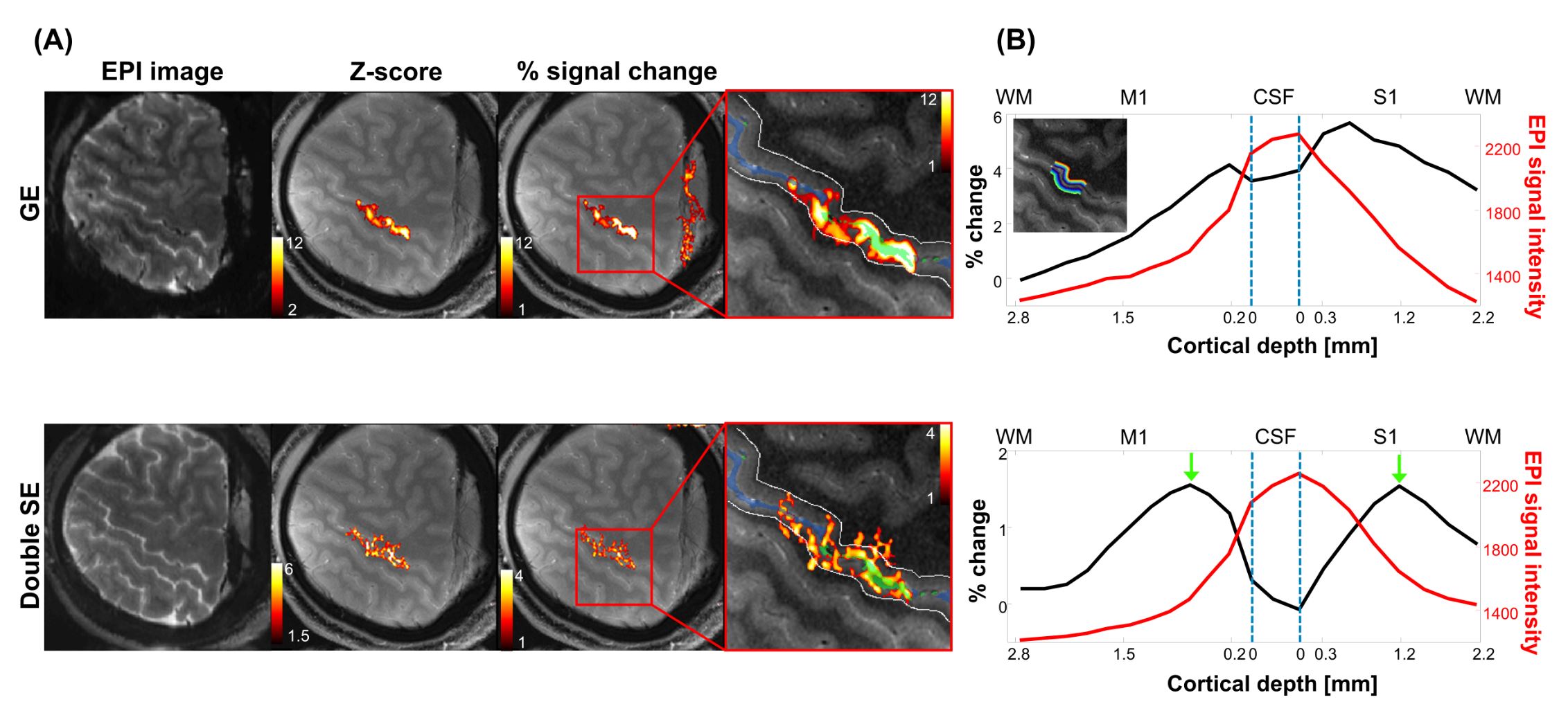
(A) EPI
images (first column), z-score maps (second column), and percent signal change
(third column) from fist clenching with touching stimulation paradigm were
shown. Red box magnifies the activated area to compare the percent signal
change pattern from dSE and GE.
(B) Percent signal
change cortical profiles from GE-EPI (upper plot) and dSE-EPI (lower plot). Red
lines are baseline signal intensity, black lines are the percent signal change.
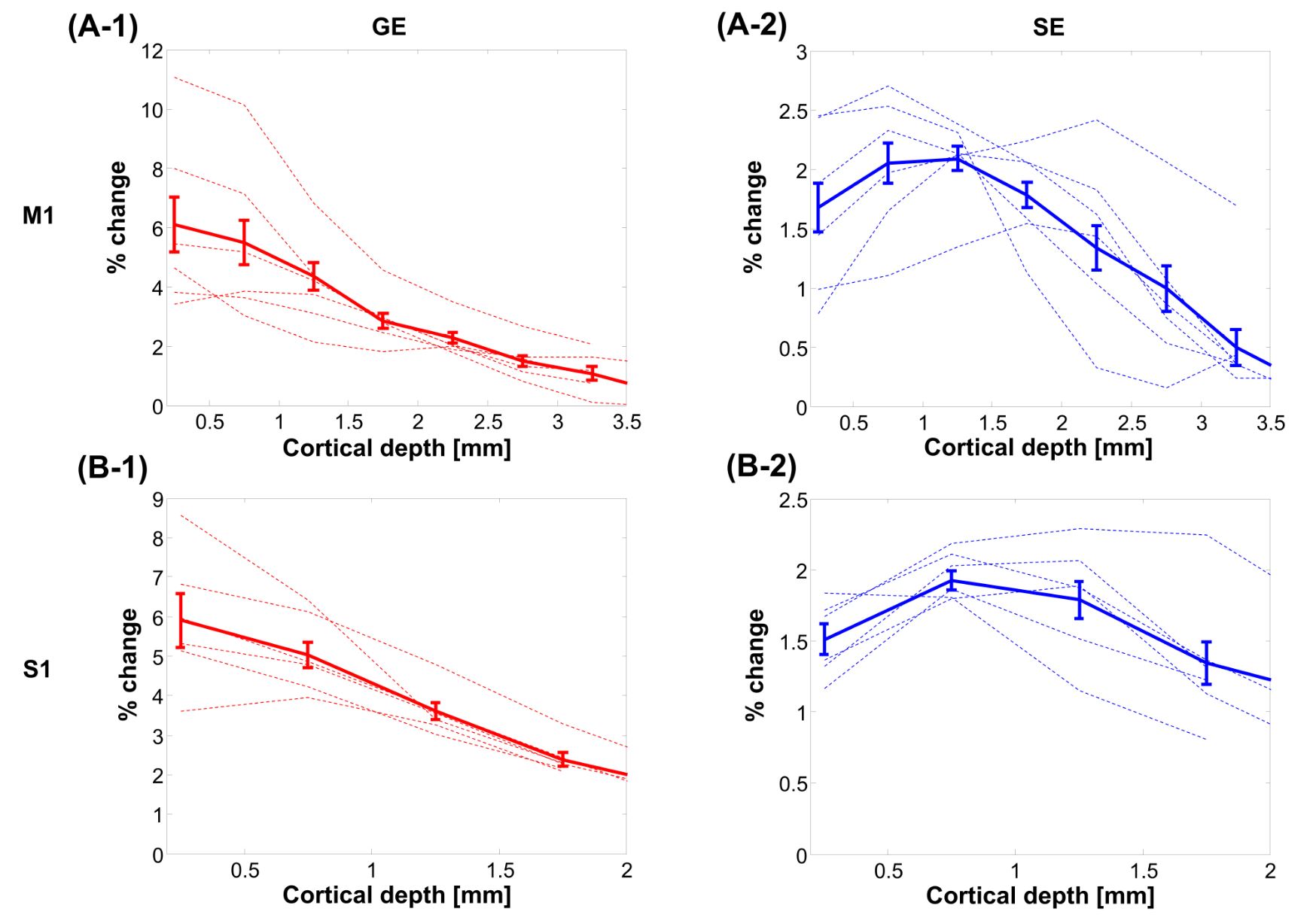
(A),
(B) Percent signal change
cortical profiles in M1 and S1. First column and second column are
corresponding to GE-EPI and SE-EPI, respectively. Error
bars on graphs are standard errors of mean (SEM)